
Live, Laugh, Lung: A review of lung POCUS for your patient in respiratory distress
Written by: Dr. Justin Varias
Edited by: Dr. Joann Hsu
It’s another busy shift.
EMS brings in a 72-year-old woman with four days of worsening shortness of breath, significantly worse overnight. She has a history of lung cancer complicated by a malignant left pleural effusion, with a left-sided PleurX catheter that is currently not draining.
She arrives tachycardic to 129, tachypneic at 28, and in obvious respiratory distress, requiring a non-rebreather. Her oxygen saturation is 98% on the NRB, but she is using accessory muscles and has decreased breath sounds bilaterally, worse on the left.
There are plenty of possibilities here.
Pneumothorax? Recurrent pleural effusion? Pneumonia? Pulmonary edema? Atelectasis?
Fortunately, we have a tool sitting right next to the stretcher.
Time to put the ultrasound probe on the chest.
Before You Scan: Pick the Right Probe
Lung ultrasound is one of those POCUS applications where the machine can look intimidating until you remember this:
You don't need fancy settings. You need the right probe and a systematic approach.
A curvilinear probe gives you a broad footprint and lower-frequency imaging, allowing you to see deeper structures. The tradeoff is that its larger footprint can make navigating between ribs a little more challenging.
A linear probe, with its higher frequency, provides excellent resolution of superficial structures—perfect for appreciating the pleural line and lung sliding.
A phased-array probe has a narrow footprint that makes it particularly useful for getting between ribs, although its lower frequency means less resolution of superficial structures.

Basics of Lung POCUS
You don't need to memorize 20+ lung ultrasound views to gather the information you need on shift.
Start with a systematic scan and know what the major findings mean.
Zone 1 corresponds to the area commonly assessed for lung sliding during the EFAST.
Zone 4 is similar to the RUQ/LUQ EFAST views, with particular attention to the diaphragm and lung base.
Zones 5 and 6 can be especially helpful when evaluating the lower lobes.

Now let's talk about the things you actually want to recognize on the screen.
A-Lines:
A-lines are equally spaced, horizontal hyperechoic lines created by reverberation artifact between two parallel strong, reflective surfaces.
In this case, between the skin/probe and the pleural line
And importantly: A-lines can be completely normal.
Seeing A-lines doesn't automatically mean "the patient has a pneumothorax."
Due to the reflective nature of the pleural lining, a portion of the ultrasound waves reflect between the pleura and skin surface multiple times, leading to the machine interpreting this as a pleural line that is further than it appears.
Since the distance between the pleura and the transducer probe/skin surface doesn't change, the artifacts are evenly spaced.
So don't panic when you see stripes!

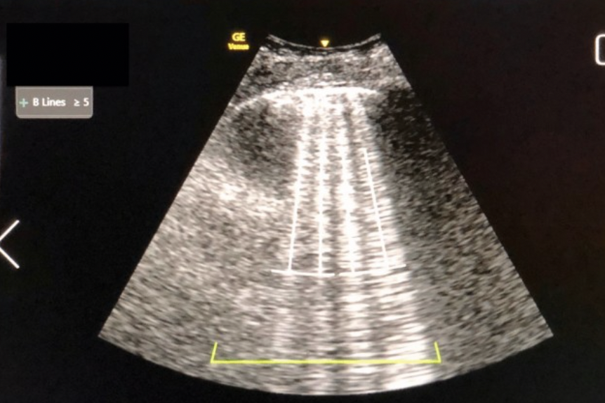
B-Lines:
B-lines look dramatically different.
They're long, vertical, hyperechoic lines that extend from the pleural line all the way to the bottom of the screen.
To evaluate for B-lines, you should increase your depth to about 10-12 cm and observe the lines extending to the far field
B-lines occur when there is increased density within the lung, which can be associated with interstitial edema, increased fluid, or fibrosis.
But don't stop there. Ask yourself, are the lines in one area or spread across multiple lung fields? This is where being familiar with your 8 main and 4 supplemental lung zones are important yet again.
Focal B-lines can point you toward a focal interstitial process or pneumonia, while diffuse bilateral B-lines can be seen with pulmonary edema, ARDS, or other diffuse interstitial disease.
C-Lines:
Subpleural consolidations can produce C-lines, appearing as a hypoechoic region with an irregular, "shredded" deep border adjacent to normal lung tissue.
It’s important to note though that C-lines can sometimes mimic a pleural effusion.
So if something looks like fluid, make sure you're actually looking at fluid—and not a consolidated piece of lung.

Lung Point: The Money Shot
When you're worried about a pneumothorax, you're looking for the lung point.
This is the point where the visceral pleura separates from the parietal pleura, and is the most specific finding for diagnosing a pneumothorax.
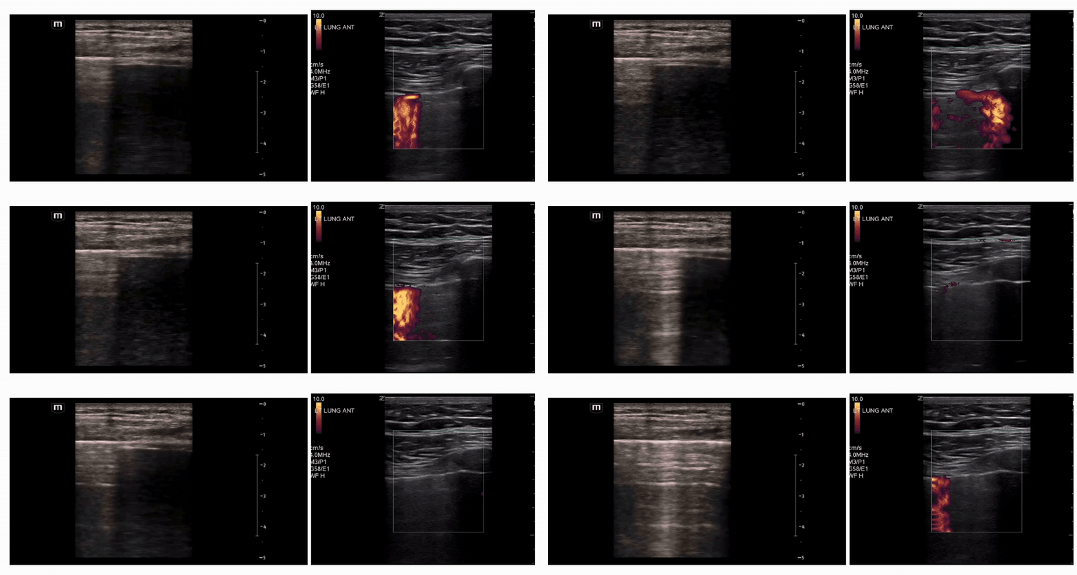
But! What if you don’t hear breath sounds on one side and you’re beginning to hallucinate whether or not you see lung sliding? Try Power Doppler!
Power Doppler can help pick up subtle movement.
If lung sliding is present, power Doppler can highlight movement along the sliding pleural line.
It may look like a curtain of fuschia/orange signal adjacent to an area of no signal at all where you know the pleura or lung tissue is sitting.
This is your lung point!
The Spine Sign:
Normally, aerated lung prevents you from seeing the thoracic spine above the diaphragm.
But when fluid occupies the pleural space, you may see a continuous hyperechoic line extending beyond the diaphragm—the classic spine sign.
This can indicate a pleural effusion or hemothorax.
For our patient, that's particularly relevant.

Hepatization:
Significant pulmonary consolidation can cause the lung to become isoechoic to the liver.
This is called hepatization. Think of it literally: the lung starts looking like the liver on ultrasound.

Air Bronchograms: Static vs. Dynamic
Once you've identified consolidated lung tissue, take a closer look for any air bronchograms.
There are two main types which can indicate different ongoing pathologies:
Static air bronchograms:
Air trapped next to consolidated lung tissue. This pattern is typically associated with conditions such as atelectasis.

Dynamic air bronchograms:
Air trapped within mucus-filled airways. These are considered pathognomonic for bacterial pneumonia.
Back to Our Patient
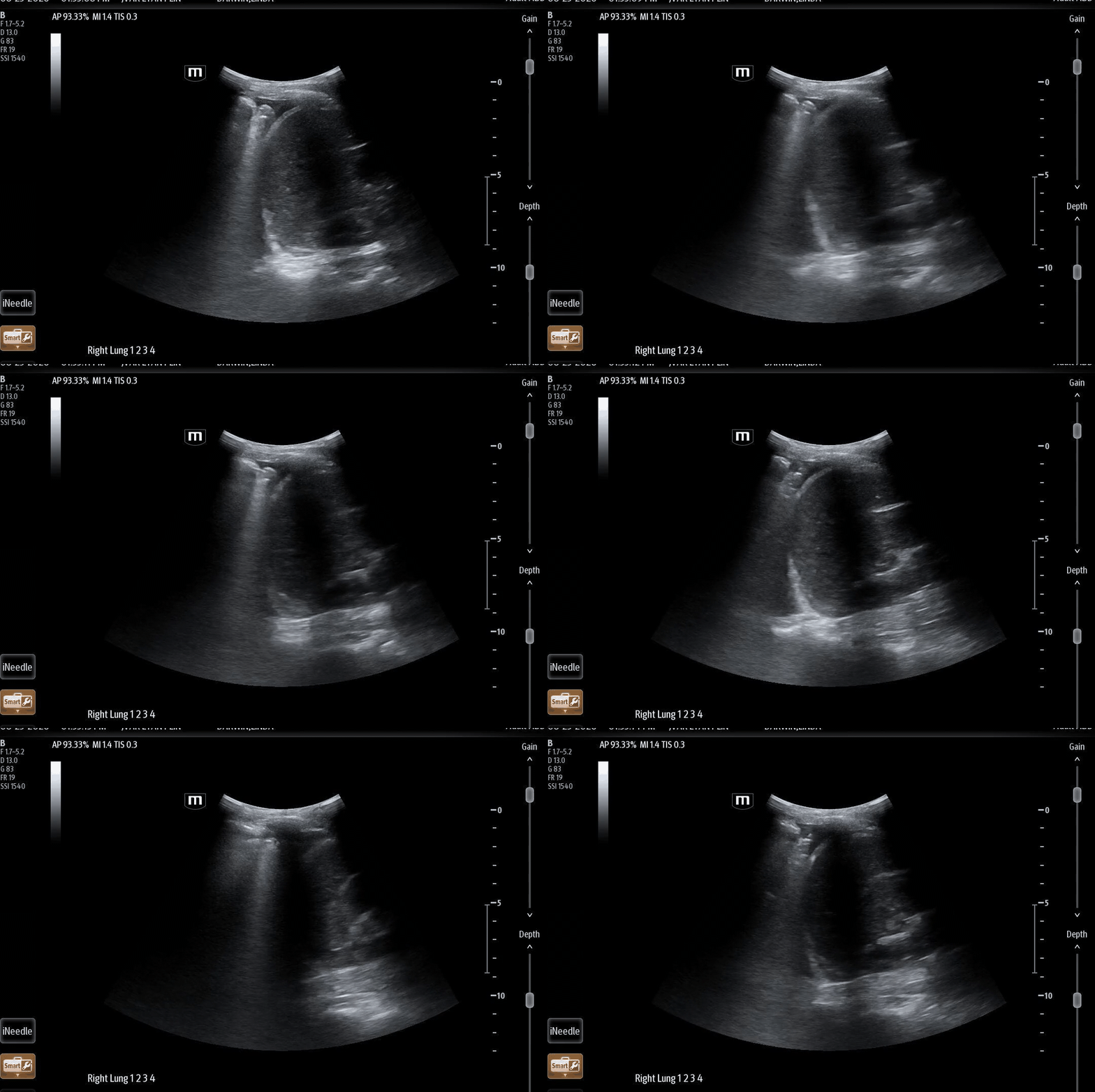
Let’s take these fundamentals of lung POCUS and apply them to our patient. This is her US, highlighting 2 main views from both the left and right apices and bases.
Right Lung:

Left lung:


We have an elderly patient with:
Known lung cancer
A malignant left pleural effusion
A left PleurX catheter that isn't draining
Worsening dyspnea
Tachycardia
Tachypnea
Respiratory distress
Decreased breath sounds, left greater than right
Knowing you have POCUS at your disposal, you can start asking more targeted bedside questions:
Is there lung sliding?
Are there B-lines?
Is there consolidation?
Do I see a lung point?
Is there an effusion?
The ultrasound isn't necessarily going to give you the entire diagnosis in one image, but it will narrow down your differential.
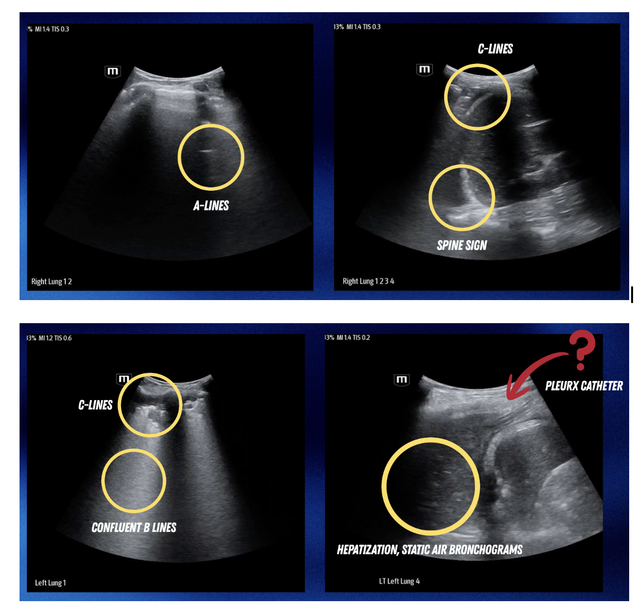
If we review her scans one more time, these are some of the main findings the team discussed at bedside:
Why This Matters in the ED
The advantage of lung POCUS isn't that it replaces your history, physical exam, chest X-ray, CT, or clinical judgment.
It's that you can perform it immediately, at the bedside, while the patient is still sick.
In a patient like ours, where the initial differential includes pneumothorax, pleural effusion, pneumonia, atelectasis, pulmonary edema, and other pulmonary pathology, lung ultrasound can quickly provide additional information to guide your clinical reasoning.
Happy scanning!
References:
https://kwaktalk.org/clinical-scan-guide#lung
https://asra.com/news-publications/asra-newsletter/newsletter-item/asra-news/2022/02/07/point-of-care-ultrasound-spotlight-lung-ultrasound
https://nephropocus.com/2019/07/01/dynamic-air-bronchograms-ultrasound-sign-of-pneumonia/
https://deepbreathe.ai/blog/lung-ultrasound-artifacts
https://www.pocus101.com/ultrasound-machine-basics-knobology-probes-and-modes/#Phased_Array_(Sector)_Ultrasound_Probe
https://www.sonosite.com/education/learn/understanding-basics-pocus-probe-types
https://nephropocus.com/2019/12/03/the-shred-sign/
https://radiopaedia.org/articles/air-bronchogram?lang=us
https://radiopaedia.org/articles/thoracic-spine-sign-ultrasound?lang=us
https://pmc.ncbi.nlm.nih.gov/articles/PMC5292837/
https://www.med.upenn.edu/POCUS/pneumothorax.html