
D Sign
Written by: Dr. Lisa Yang
Edited by: Dr. Joann Hsu
The case:
86yoM presenting with worsening short of breath that prevents him from lying down and decreased exercise tolerance. He has a past medical history of CAD s/p stent (2019), HFpEF, rheumatic mitral valve s/p replacement, single lead pacemaker, HTN, and HLD. His vitals are stable and his pulse ox is 96% on room air, but his physical exam reveals decreased breath sounds.
Now, before we get into our patient’s echo, let’s discuss what we usually want to evaluate on an echo.
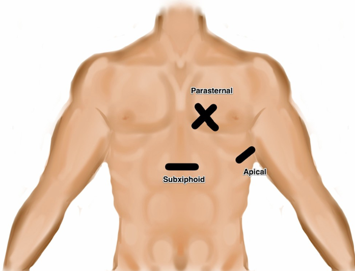
The views of an echo are relatively straight forward—four views as follows: parasternal long, parasternal short, apical four chamber, and subxiphoid.
Using these four views, we must develop an overall assessment of the patient’s cardiac function.
There are many systematic ways this is taught; for example, going through the five Es (effusion, ejection, equality, exit, entrance).
What matters for you clinically is choose a method that allows you to systemically perform and interpret an echo to answer your relevant questions in the ED.
Today let’s walk through one method I have named “follow the flow”.
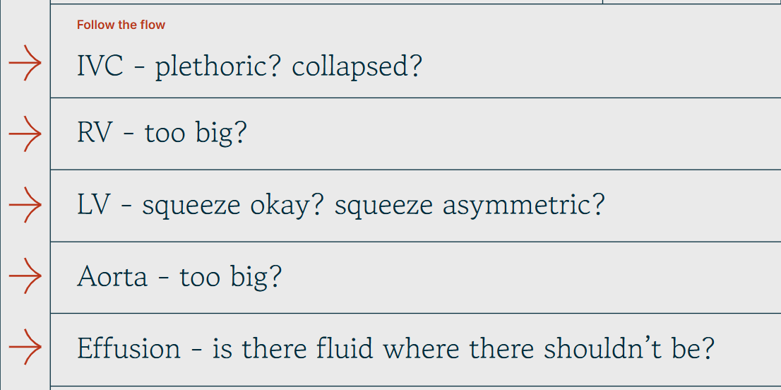
We start with the blood entering the heart through the IVC.
As always in POCUS, we want to answer a binary question.
Our questions here are: “Is the IVC plethoric? Is it collapsed?”
A plethoric IVC may indicate a fluid overload state for example, or a sign of obstructive shock
Next, we enter the right side of the heart.
We ask ourselves: “Does the RV look too big?” - which would be concerning for right heart strain.
After the right heart comes the left heart.
“Does the EF look reduced? Is there wall motion abnormality?”
Now we exit the heart: “Is the aortic root dilated?”
Finally —”In all my views, did I consistently see fluid around the heart?” - concerning for a pericardial effusion.
Instead of trying to remember what each E stands for, simply remembering the path of blood throughout the heart can make sure you answer all your clinical questions.
Now that we have the basics covered, let’s return to our patient and look at his echo.
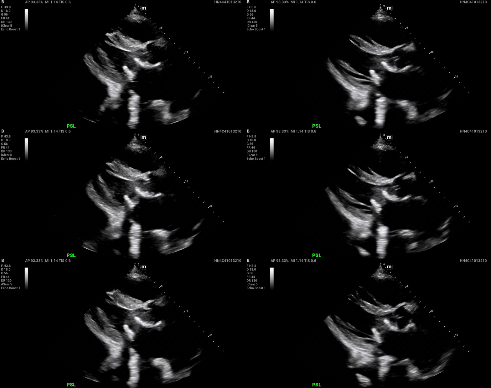
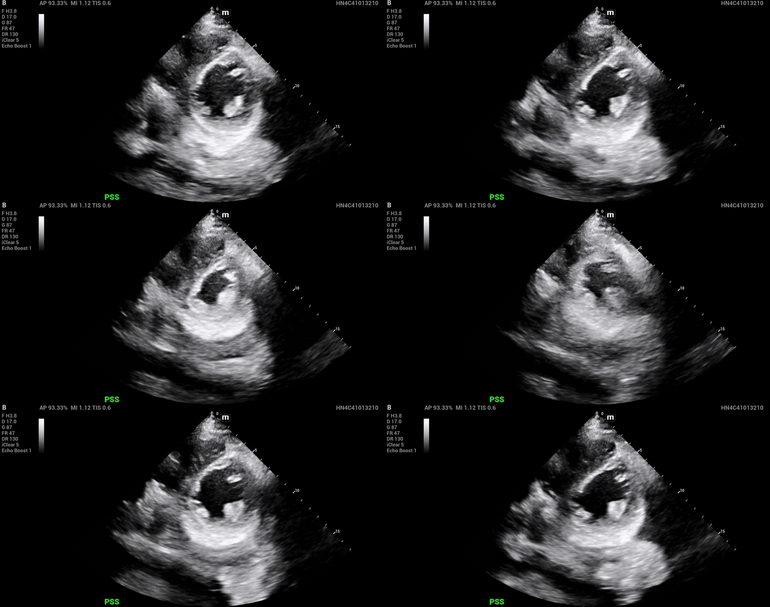
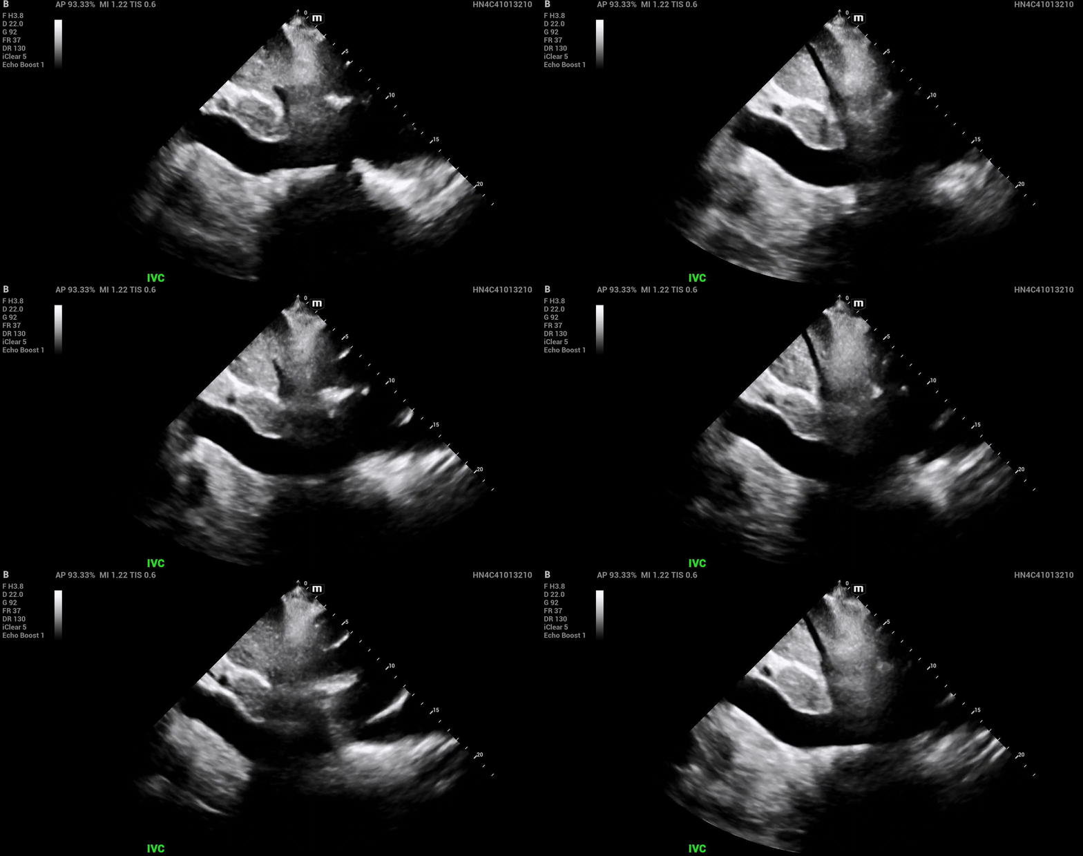
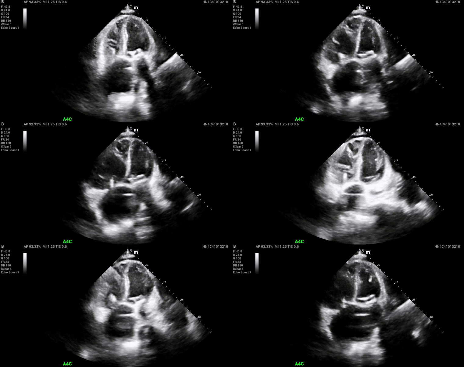
From these views, let’s draw our conclusion using our template from above.
Plethoric IVC
Enlarged RV
Preserved EF
Septal bowing (D sign)
Normal sized aortic root
These findings are concerning for right heart strain.
We are going to dive into POCUS findings that suggest right heart strain, specifically D-sign, or bowing of the interventricular septum.
There are two main underlying causes of right heart strain that can be differentiated using the D-sign: pressure overload and volume overload.
Pressure overload is associated generally with primary lung pathology. This includes pulmonary embolism, pulmonary hypertension, and ARDS.
Volume overload, on the other hand, is associated with etiologies such as severe tricuspid regurgitation, decompensated heart failure, or iatrogenic causes such as aggressive volume resuscitation.
And the great thing is, these two types of overload look completely different on ultrasound.
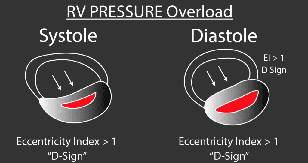
In pressure overload, D sign is going to be seen throughout the cycle, but most pronounced during systole.
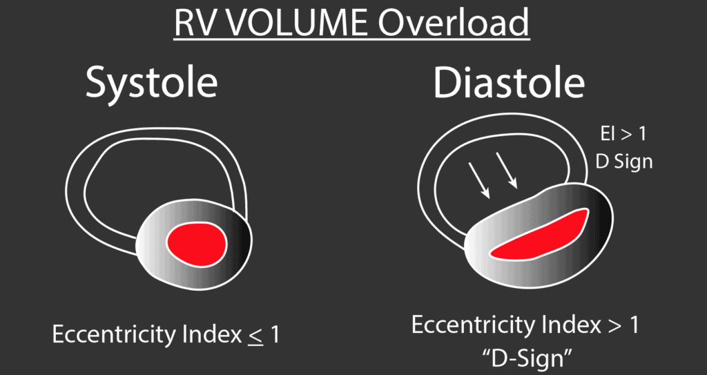
In volume overload, D-sign is going to be most pronounced during diastole, and essentially resolved during systole.
A study published in the Journal of the American College of Cardiology recruited 33 patients—11 with normal physiology, 11 with primary pulmonary hypertension, and 11 with severe tricuspid regurgitation due to tricuspid valve resection. -
Echocardiograms of these patients corroborated this teaching, writing, “Right ventricular systolic overload in primary pulmonary hypertension resulted in substantial leftword ventricular septal shift that was most marked at end-systole and early diastole and decreased substantially by end-diastole. Right ventricular diastolic overload after tricuspid valve resection resulted in maximal leftward ventricular septal shift at end-diastole sparing end-systole and early diastole.”
But does that mean there’s another ultrasound measurement you have to memorize now? By no means! Let’s go over why this makes perfect physiologic sense.
In RV pressure overload, the RV is pumping against increased afterload due to a primary pulmonary pathology.
Therefore, it can be thought of as a force coming from the direction of the pulmonic valve.
The pulmonic valve is open during systole, allowing this pressure to confront the right ventricle.
And guess what, the D-sign is most pronounced during systole in pressure overload.
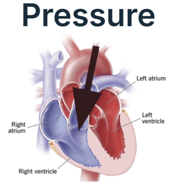
Likewise, in RV volume overload, the main pathology is due to overfilling of the RV.
The RV fills in diastole, and empties during systole.
Therefore, it follows that the D-sign is most pronounced during diastole, and nearly resolved in systole.
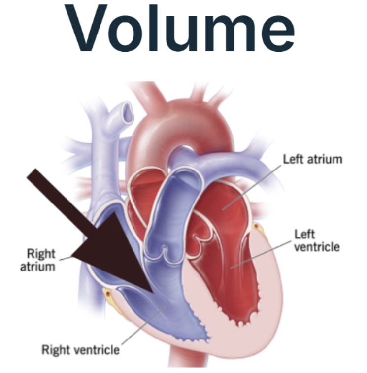
Now that we are armed with this new knowledge of interpreting the D-sign, let’s return to our patient’s parasternal short axis view—this time, with some stills to help us.
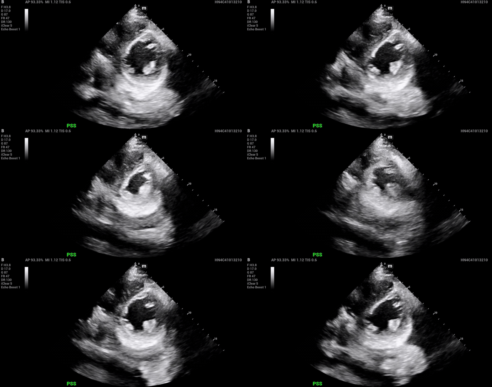
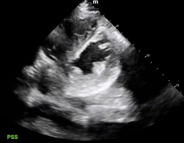
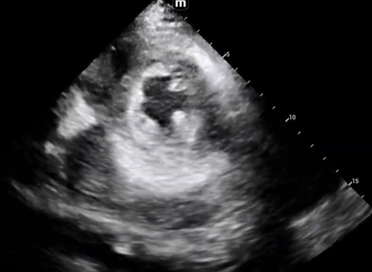
What we are seeing here is a D-sign during diastole, with near resolution during systole. And what does that mean folks? Volume overload.
Let’s return back to the etiologies we talked about.
Severe tricuspid regurgitation
Decompensated heart failure
Iatrogenic aggressive volume resuscitation.
Given that this patient has just presented, we can focus more on the first two items.
Our patient has a pacemaker in his right ventricle, could this be causing some regurgitation? Definitely.
Our patient also has a diagnosis of HFpEF, could this have now evolved into HFrEF? Definitely.
Our patient is experiencing volume overload and not RV pressure overload, is he suffering from a massive pulmonary embolism? Less likely, given all the new information we have gained.
So what happened to our lovely patient?
His ED workup included a CXR demonstrating a left pleural effusion, a CTPE that demonstrated no embolisms, and non-actionable troponins and BNPs. He was admitted for workup, and found to have a preserved ejection fraction, “torrential tricuspid regurgitation”, and bioprosthetic mitral valve prolapse with regurgitation.
In summary, what does this mean for our clinical practice?
If a patient presents with acute dyspnea and a D-sign on ultrasound that looks more like volume overload than pressure overload, can we skip the CTPE? No. Remember that POCUS is just one of many tools we use in addition to history, physical exam, vitals, etc. But it can help guide our workup and strengthen certain differential diagnoses.
In this patient, who was already on a blood thinner for his prosthetic valve, saturating well on room air, and presenting with his specific risk factors and volume overload on echo, it can help prioritize the differential with more finesse.
Happy scanning!
References
Dinh, V. (no date) The D sign - right heart strain from pressure vs volume overload, POCUS 101. Available at: https://www.pocus101.com/the-d-sign-right-heart-strain-from-pressure-vs-volume-overload/ (Accessed: 06 September 2026).
Louie, E, Rich, S, Levitsky, S. et al. Doppler echocardiographic demonstration of the differential effects of right ventricular pressure and volume overload on left ventricular geometry and filling. JACC. 1992 Jan, 19 (1) 84–90.
https://doi.org/10.1016/0735-1097(92)90056-S
Ryan, T, Petrovic, O, Dillon, J. et al. An echocardiographic index for separation of right ventricular volume and pressure overload. JACC. 1985 Apr, 5 (4) 918–924.
https://doi.org/10.1016/S0735-1097(85)80433-2
Weerakkody Y, Elfeky M, Sharma R, et al. Right heart strain. Reference article, Radiopaedia.org (Accessed on 06 Sep 2026) https://doi.org/10.53347/rID-31600