Lower abdominal mass on POCUS
Written by: Dr. Callie Winters
Edited by: Dr. Joann Hsu
Case:
An 81-year-old patient presents to the ED from nursing home with complaint of abdominal distension and hematuria. The patient was sent in by an outpatient provider to rule out SBO.
PE: palpable LLQ mass that is nonmobile, tender, nonfluctuant, with no overlying skin changes
(+) chronic indwelling foley w/ blood at theurethral meatus
Rest of exam unremarkable
You place your probe on the palpable mass:
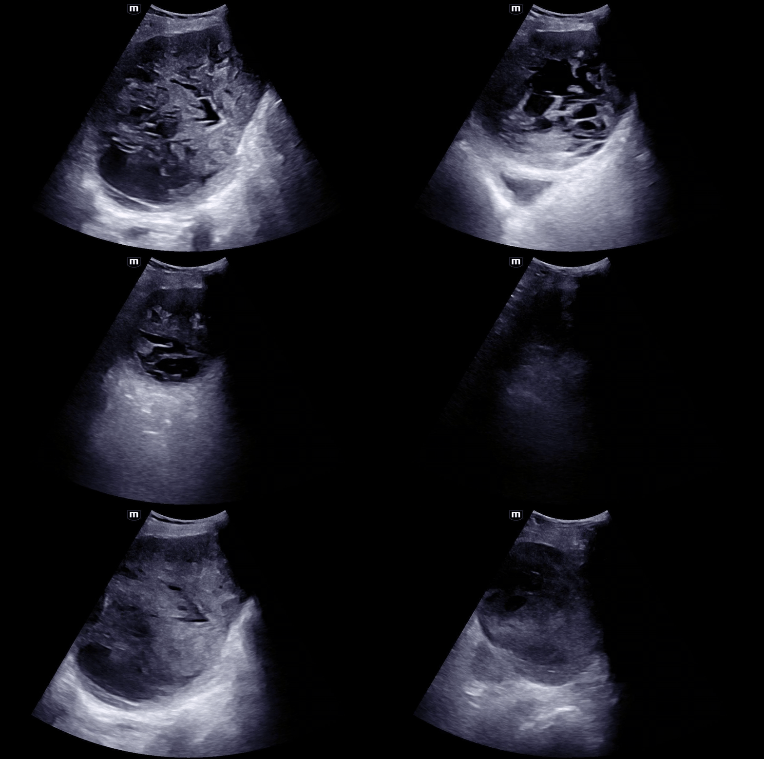
You see a very large, complex mass. No obvious bowel or peristalsis. You can see a glimpse of the bladder at the farfield.
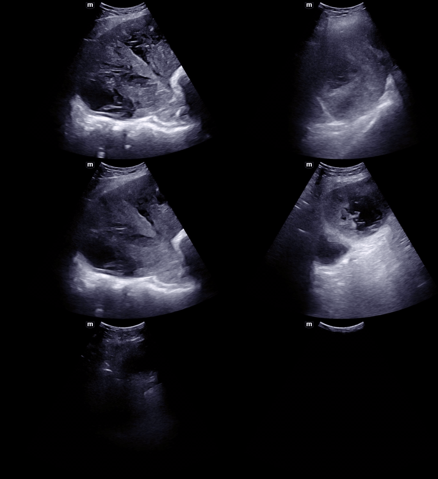
Here it is from another axis.
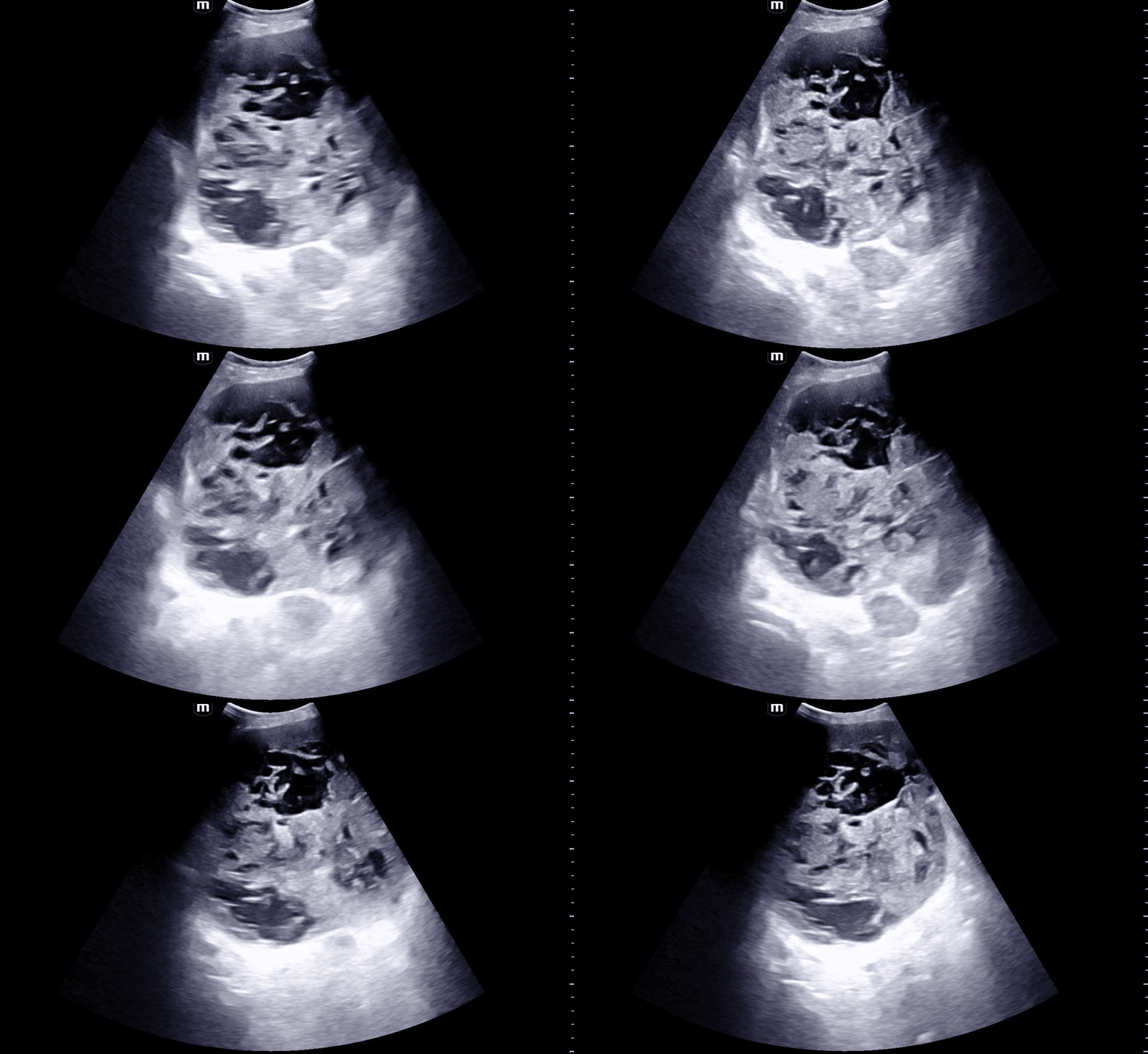
Here is the same mass with external pressure from the probe, to see if we can elicit any internal movement of contents, like we would see in a fluid filled complex structure such as an abscess. As you see here - there is no internal movement of contents.
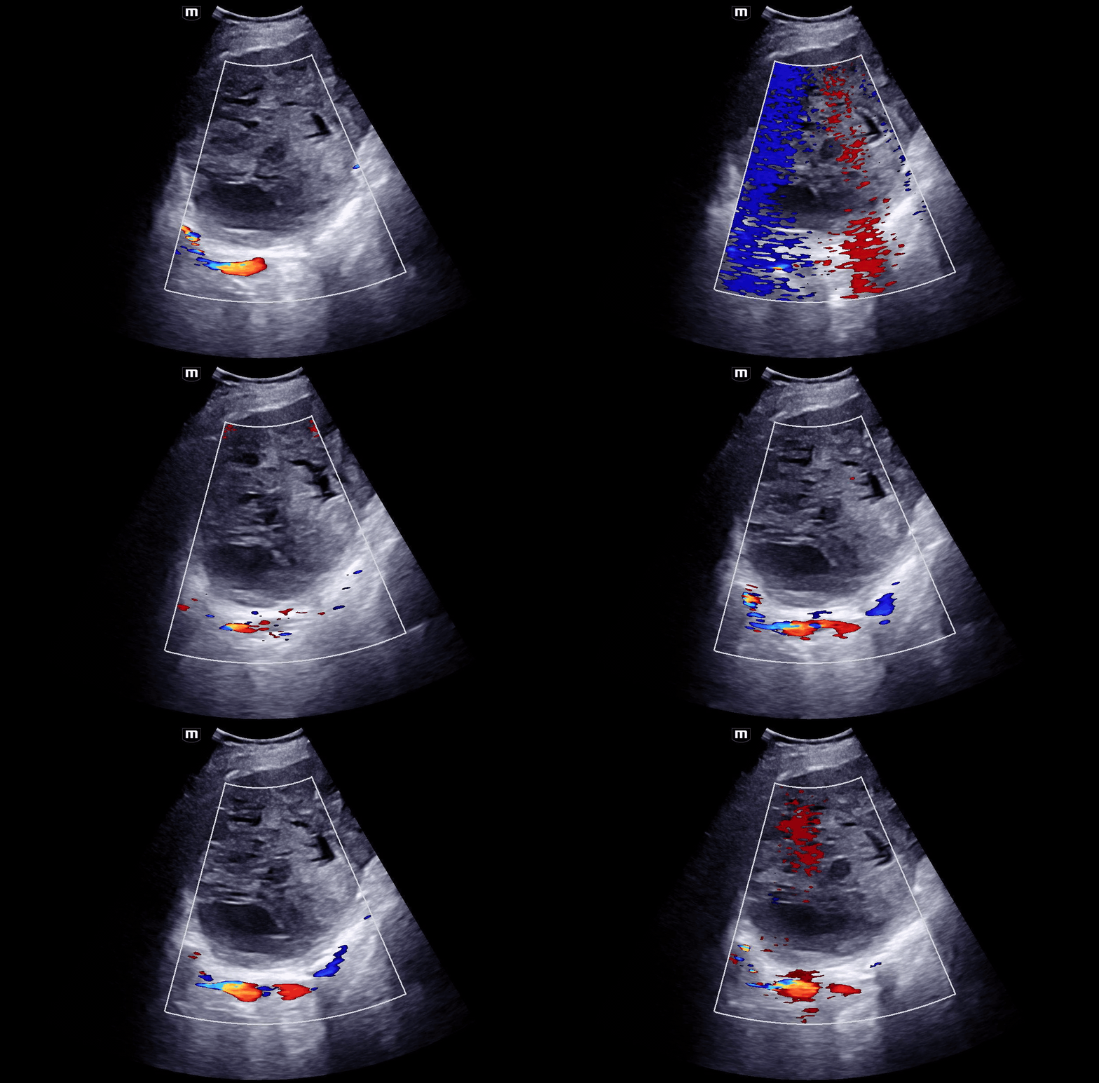
There is no significant internal flow in this structure.
Not shown here: bilateral hydronephrosis on bedside ultrasound.
Distinguishing solid masses from other structures on POCUS
Is it an abscess?
An abscess could have:
Anechoic/hypoechoic areas
Though unlike a simple cyst, may containhyperechoic debris
Swirling of internal debris w/ compression"pusastalsis"
*maybe* septations
Could have dirty shadowing/air
Increased posterior acoustic enhacement as is found in a fluid filled structure
Encasement/defined rind or “contained”
Surroundingcellulitis/edema/cobblestoning
Systemic signs/symptoms of infection, clinical correlation is important!!
An abscess should not have:
Significant internal vascularity (color flow)
The appearance of complex, fixed, solid structure - can be hard to differentiate
Is it bowel?
Bowel could have:
Peristalsis (withoutcompression)
If SBO:
to-and-fro or potentially absent peristalsis
Plicae circulares/keyboard sign visible
Dilated loop of small bowel greater than or equal to 2.5 mm
Air + fluid
Plicae circulares (SB) orhaustra (LB)
Continuity w/ rest of bowel
Bowel should not have:
Internal septations/complexinternal structure
It should be poop in there
Internal vascular flow
Could it be a solid soft tissue mass?
A soft tissue mass could have:
Complex/hyperechoic/mixedechogenic internal structure
May resemble functional solid organ tissue
Internal vascular Flow
Well circumscribed borders
A soft tissue mass should not have:
Significant compressibility or fluctuance
No “swirl sign” - when liquid internal contents swirl during compression as is seen in an abscess
Peristalsis
Bowel loops or stool
Prostatic Leiomyomata
A rare benign tumor of the prostate gland
2023 case report cited fewer than 50 cases ever reported
Diagnosed via histology
Must be distinguished from malignancy
Can present with urinary retention
Well-circumscribed, non-invasive, with amultiloculated, cystic cut surface
Treatment options:
Surgical excision: prostatectomy,transurethral resection
Prostatic artery embolization, observation ifasymptomatic
Case conclusion:
CTAP revealed:
foley balloon malpositioning in the prostatic urethra
18.5 cm pelvic mass (increased size from prior)
bilateral hydronephrosis
Admitted for AKI
Patient refused surgery for leiomyoma
AKI improved following foley replacement
Discharged back to nursing home after one week in hospital
Takeaways
In rare cases, male urinary retention canbe due to prostatic leiomyomata rather than simple BPH
Treatable, benign tumors
Dx=histology -> must be differentiated from malignancy
To differentiate solid masses from other structures
Use compression
Use color-flow
Use common sense: don't needle aspirate/I+D if unsure
Happy scanning!
References: