
When Milk Won’t Pass
Written by: Dr. Khin Oo
Edited by: Dr. Joann Hsu
Case:
A 5 week old female ex-full term, twin birth, presenting with vomiting and diarrhea for the past few days.
Patient’s mother described that the diarrhea continued and despite giving her formula or Pedialyte, 2 oz every 2 hours, and the baby vomited every single time, a large amount. Patient was not tolerating PO today. Last urine was this morning. The patient was described to be hungry after each vomit.
Physical exam is unremarkable.
Vitals:
BP 96/46 | HR 144 | T 37.4 C | SpO2 100% RA
Using a linear ultrasound probe a flat footprint to create high-resolution images of shallow or surface-level structures in children, let’s take a look at what we saw on the epigastric and RUQ region in transverse and sagittal views:
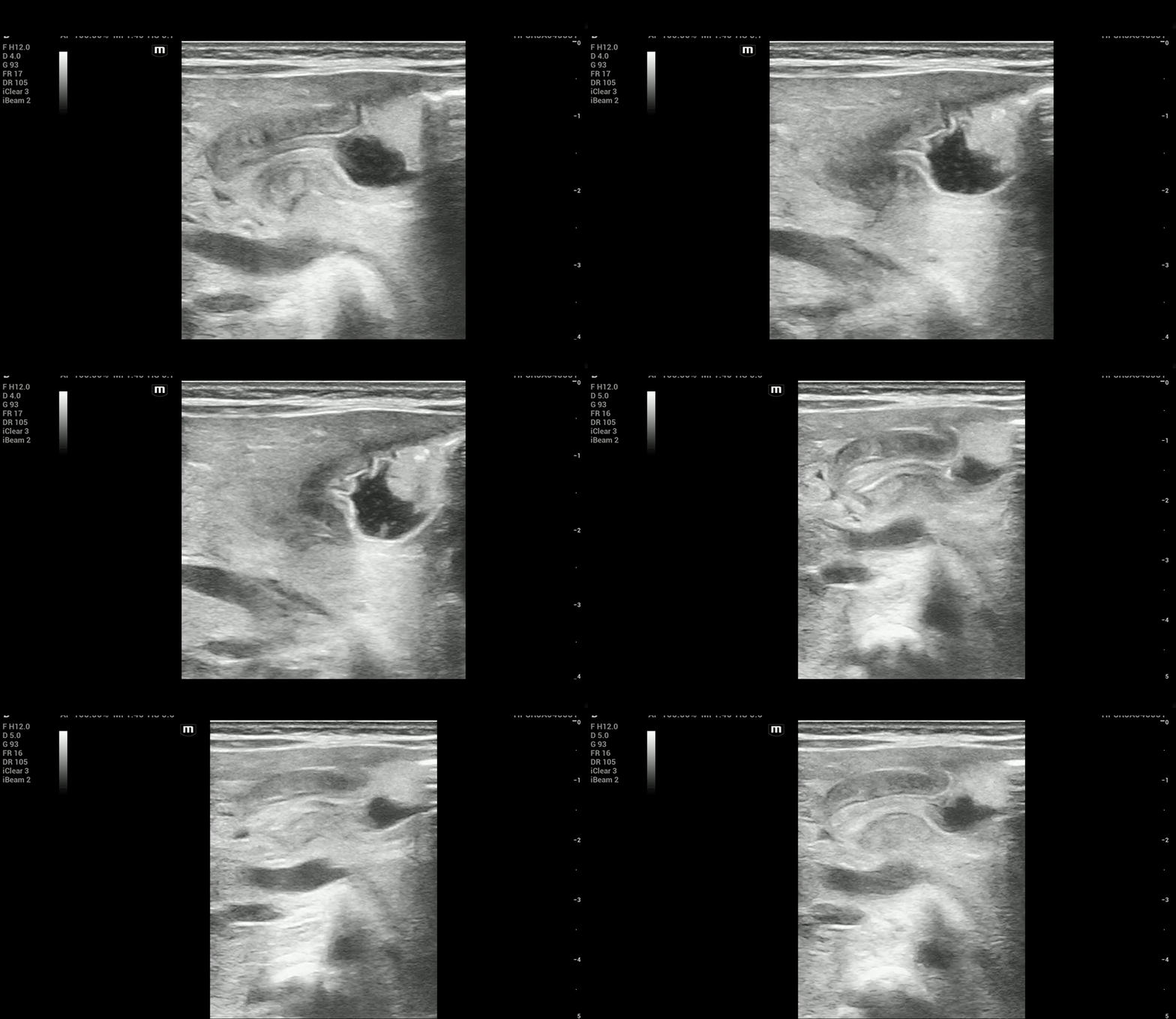
Figure 1: sagittal view
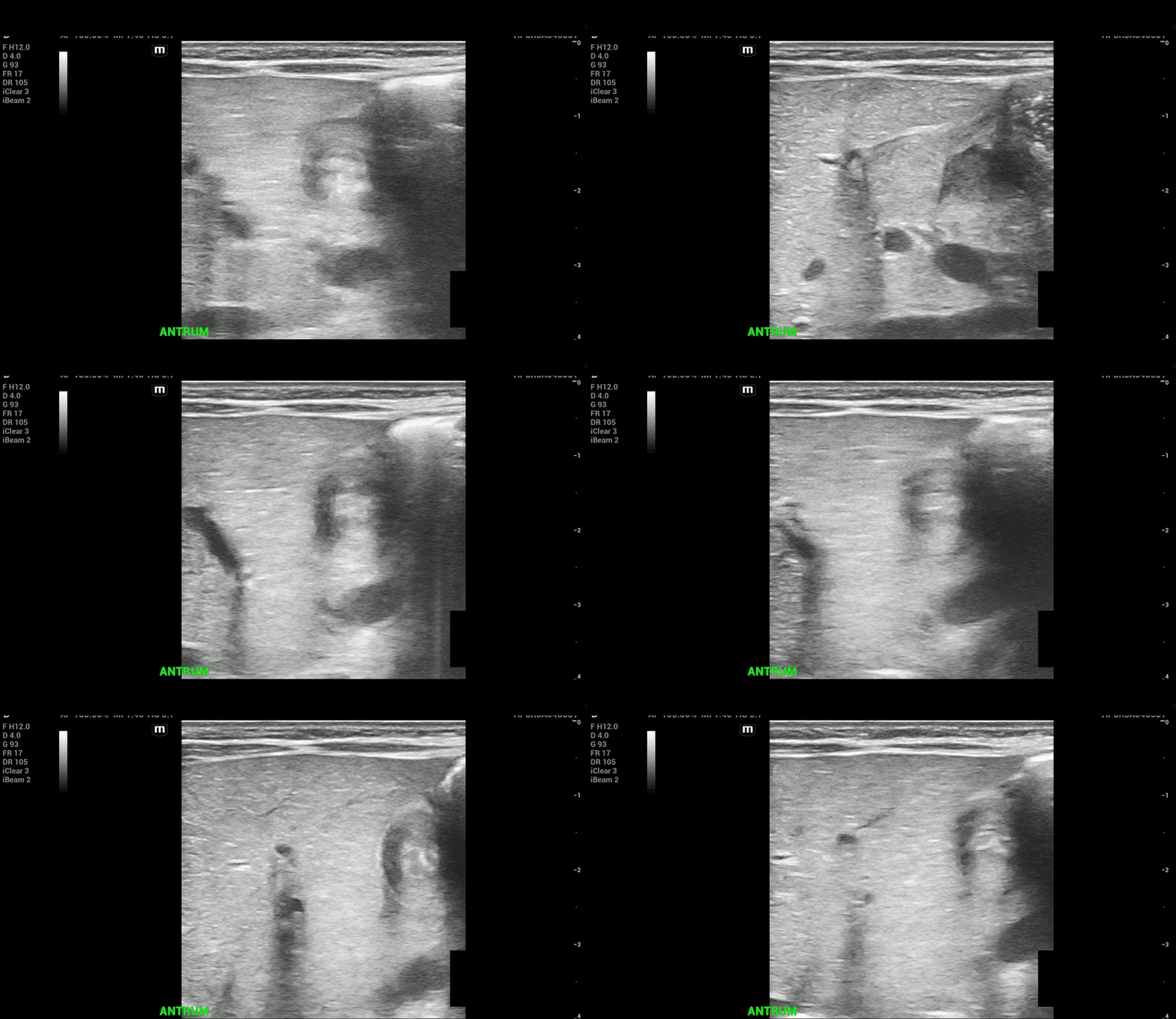
Figure 2: transverse view
What do you see?
You see the fluid-filled antrum of the stomach and following it distally, you see the pylorus.
Long axis view of the pyloric muscle highlighted by arrows at the distal end of the stomach
In between the thickened hypoechoic pyloric muscles (muscularis externa) is the hyperechoic pyloric channel made up of gastric mucosa.
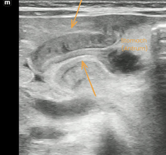
Figure 3: Hypertrophied pyloric muscle in long axis
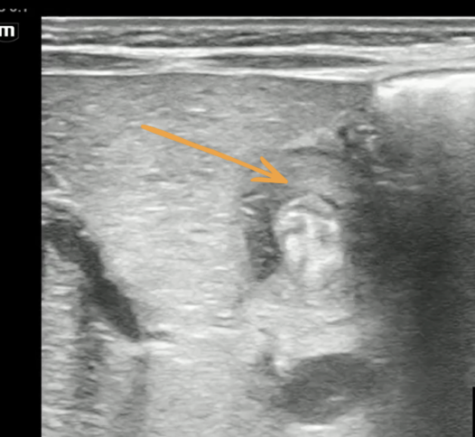
Figure 4: Hypertrophied pyloric muscle in short axis
Tip: Identify known landmarks if you have trouble locating the pylorus
Find the gallbladder adjacent to the medial edge of the liver and look even more medially to locate the pylorus.
Alternatively, find the fluid-filled stomach and follow it distally to the pylorus.
Use the aorta/ superior mesenteric artery as additional landmarks if necessary (pylorus is anterior to them).
Pyloric Muscle Thickness
Thickness of the pyloric muscle can be measured in the long or short axis view
Pyloric muscle thickness, i.e. diameter of a single muscular wall (hypoechoic component)
Hypertrophied pyloric muscle in short axis measuring 3.8 mm
A thickness of 3 mm is considered abnormal or hypertrophied
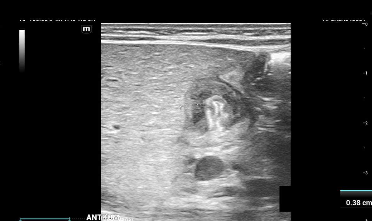
Figure 5: Hypertrophied pyloric muscle in short axis measuring 3.8 mm
Pyloric Transverse Diameter
Pyloric transverse diameter measuring 13.8 mm
Pyloric transverse diameter >14 mm considered abnormal or hypertrophied
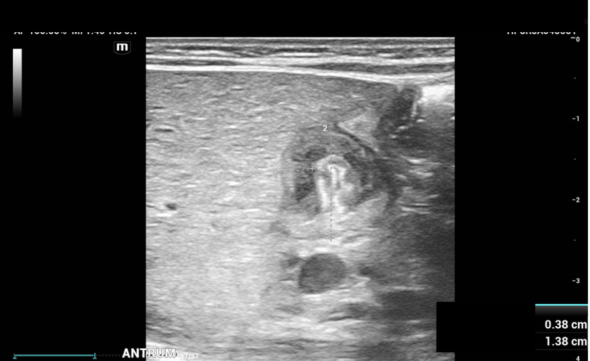
Figure 5: Pyloric diameter in short axis measuring 13.8 mm
Pyloric Channel
Pyloric channel measuring 19.1 mm
Length, i.e. longitudinal measurement of >15-17 mm considered abnormal or elongated
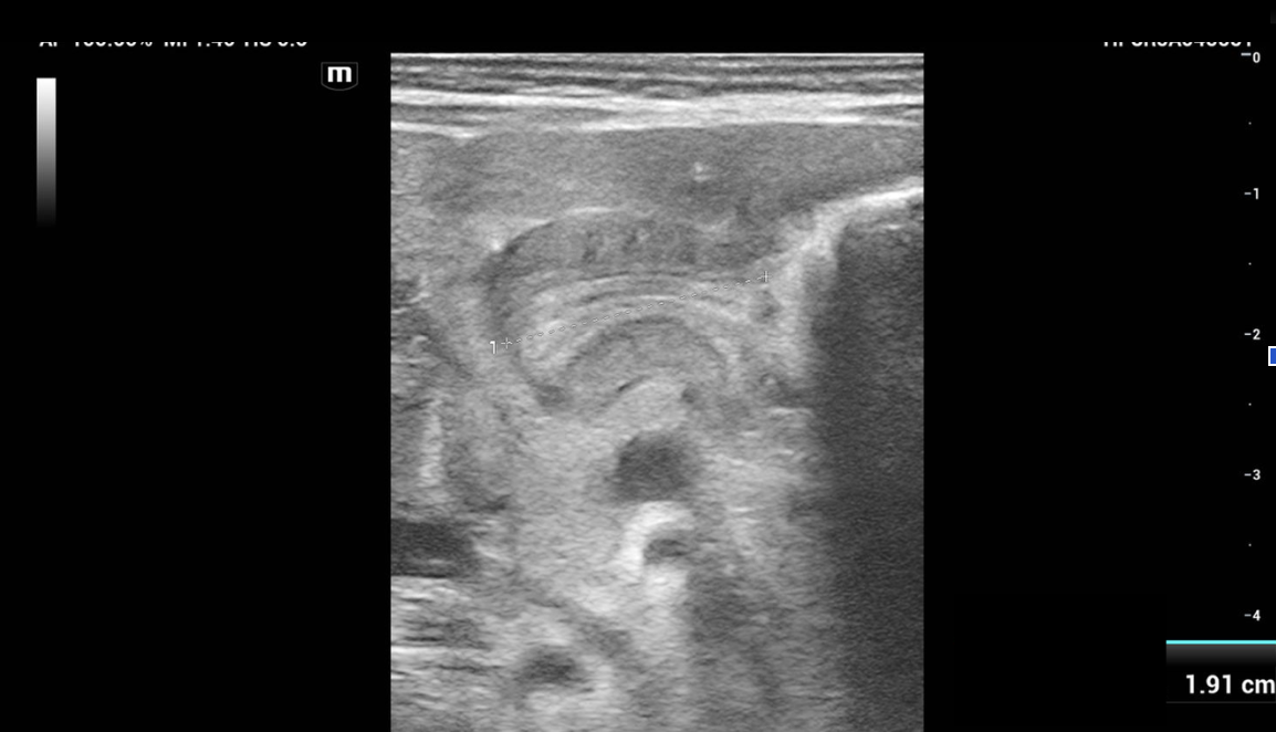
Figure 6: Elongated Pyloric channel in long axis 19.1 mm
The "Pi" Breakdown (π = 3.1415) :
3 — Pyloric muscle thickness: Greater than 3 mm (measured as a single muscular wall on a transverse ultrasound image).
14 — Transverse diameter: Greater than or equal to 14 mm (wall-to-wall transverse measurement).
15 — Pyloric length: Greater than 15 mm (longitudinal channel measurement, sometimes noted up to 15–17 mm
Based on the patient’s ultrasound findings, the appearance is consistent with a pyloric stenosis
Key Sonographic Signs
Target Sign - echogenic mucosa surrounding thickened pyloric hypoechoic muscle
Antral nipple sign- redundant pyloric mucosa protruding into the gastric antrum
Cervix sign - indentation of the elongated, narrow pylorus canal into the fluid-filled antrum. The most telling sign, however, was that no gastric contents were seen to pass through the pylorus at any time during the examination.
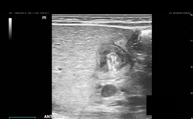
Figure 7: Target Sign
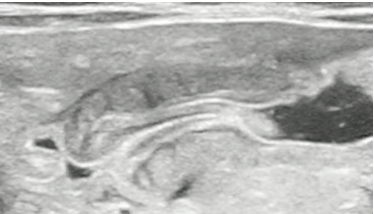
Figure 8: Antral Nipple Sign
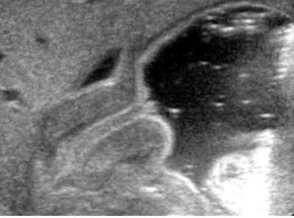
Figure 9: Cervix Sign
Pyloric Stenosis
Hypertrophic pyloric stenosis is an idiopathic, acquired condition generally presenting at 2-12 weeks of age.
Caused by hypertrophy of the circular muscle of the pylorus, causing gastric outlet obstruction.
Classic findings: projectile non-bilious vomiting, palpable “olive” at the epigastrium, and hypochloremic metabolic alkalosis in bloodwork
Ultrasound is the gold standard in diagnosis, with sensitivity 97-100% and specificity 99-100%
Initial medical management is essential with rehydration and correction of electrolyte imbalances. This should be completed before surgical intervention.
Treatment is surgical with a pyloromyotomy in which the pyloric muscle is divided down to the submucosa. This can be performed both open and laparoscopically. The operation is curative.
Back to the Case
US shows pyloric channel is thickened, measuring up to 5 mm with an elongated channel measuring up to 1.8 cm. Gastric contents are not seen passing through the pylorus.
Surgery was consulted.
The patient was admitted PIMU under the surgical service with for operative treatment
Started the patient on 1 x M IV fluids
Take Home Points
Use π = 3.1415, 3 for Pyloric muscle thickness, 14 for Transverse diameter and 15 for Pyloric length
Hypertrophic pyloric stenosis is an idiopathic, acquired condition generally presenting at 2-12 weeks of age.
Try feeding the child and follow the stomach contents as it moves through the pylorus
Happy scanning!
References
https://www.acep.org/sonoguide/advanced/pediatric-pyloric-stenosis
https://radiopaedia.org/cases/infantile-hypertrophic-pyloric-stenosis
https://radiopaedia.org/articles/pyloric-stenosis-1?lang=us
https://radiopaedia.org/articles/cervix-sign-pyloric-stenosis?lang=us
https://radiopaedia.org/cases/pyloric-stenosis#image-207