
A Sea of Lungs
Written by: Dr. Megan Wong
Edited by: Dr. Joann Hsu
A 63-year-old male with HTN presents with 2 weeks of worsening dyspnea on exertion and chest pain. He was referred by his PCP for concern of pulmonary embolism given an elevated outpatient D-dimer and left lower extremity edema. He denies a history of smoking, previous blood clots, fever, chills, upper respiratory infection, or recent weight loss.
The physical exam showed decreased breath sounds at the left lung base and good air entry throughout the right lung. He also had trace left lower extremity edema. His vitals were initially within normal limits, but after reexamination he became hypoxic to 92% and was put on 2L nasal cannula.
Given his worsening dyspnea, we used a curvilinear probe to take a look at his lungs, starting with the left lung base.
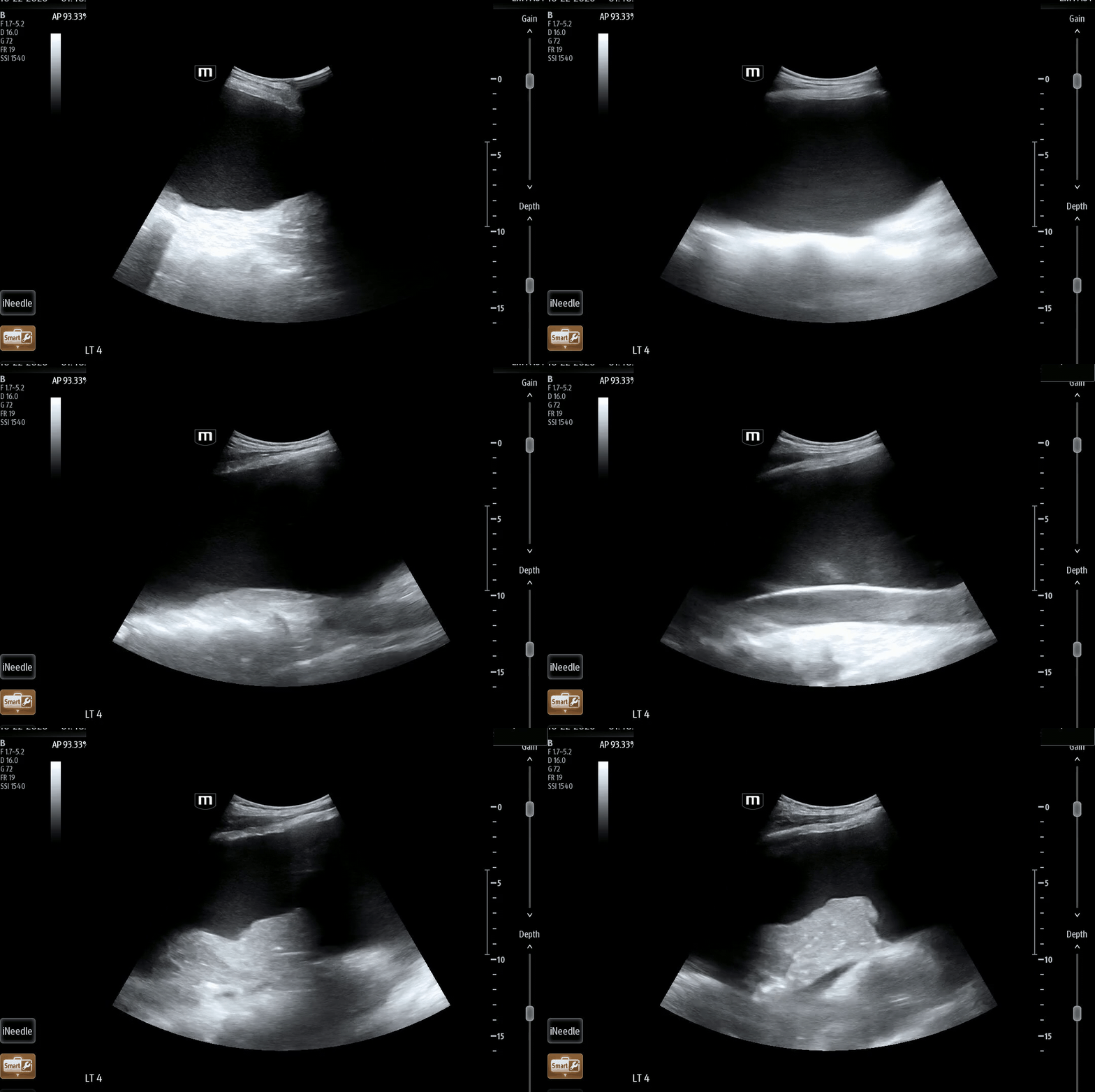
Figure 1: Left lung zone 4
What do you see?
Above the diaphragm there is a large, dark (anechoic) collection of fluid. Making its appearance towards the end of the clip, the lung is no longer sitting against the chest wall and appears to be ‘floating’ in a sea of fluid. This is a pleural effusion.
Point of care ultrasound (POCUS) is over 90% sensitive and specific for detecting pleural effusions. Now we’ll explore two main questions:
What ultrasound findings are suggestive of pleural effusions?
Can we use POCUS to tell whether the effusion is made up of transudative or exudative fluid?
Ultrasound Findings
First, let’s analyze a scan of normal lungs at zone 4:
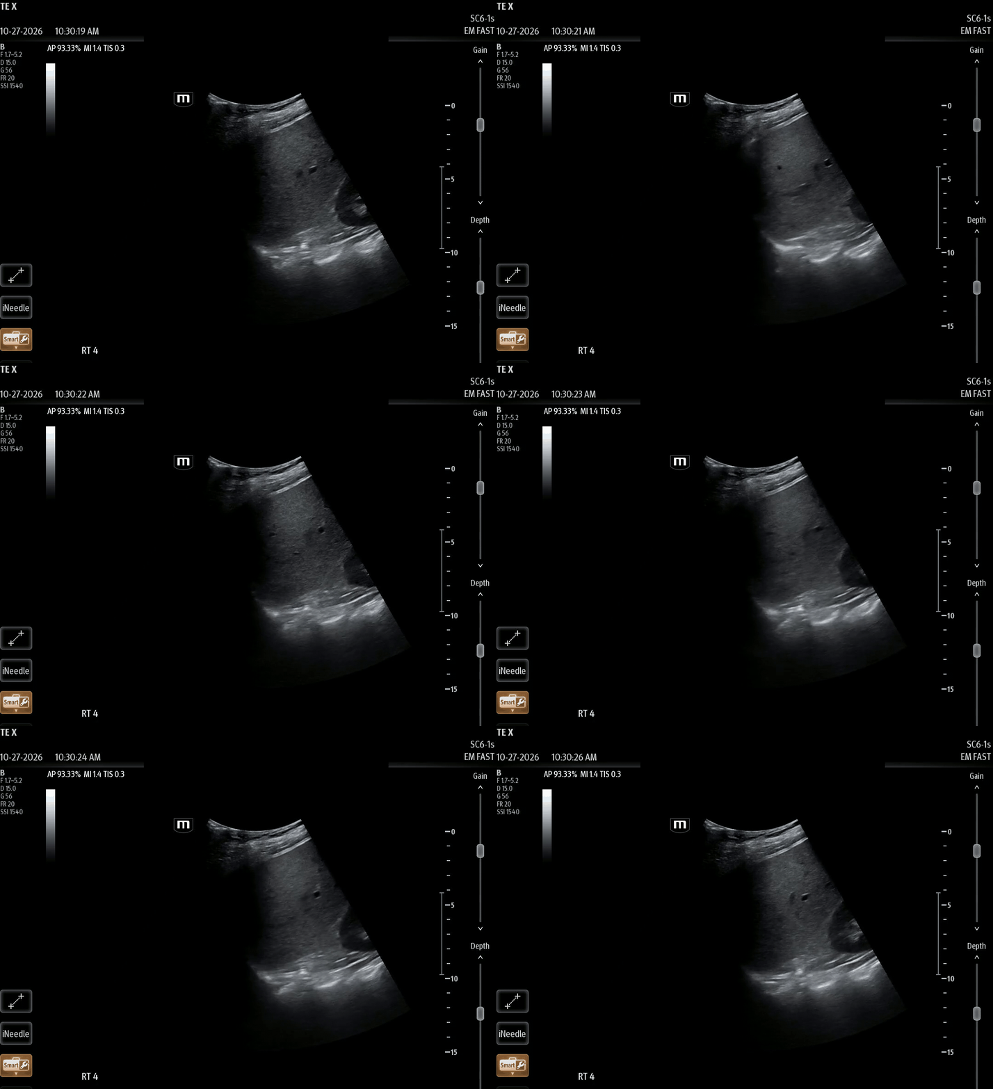
Figure 2: Normal Right lung zone 4
On a normal well-aerated lung, air blocks and scatters ultrasound waves so the farfield thoracic spine does is not visible above the diaphragm, so it is not seen at all above the diaphragm. The air also causes a dark ‘curtain’ to open and close as the patient inhales and exhales.
On inhalation, the lung moves caudally/ curtain closes towards the right of the screen.
On exhalation, the lung moves cephalad/ curtain opens towards the left of the screen.
Spine sign
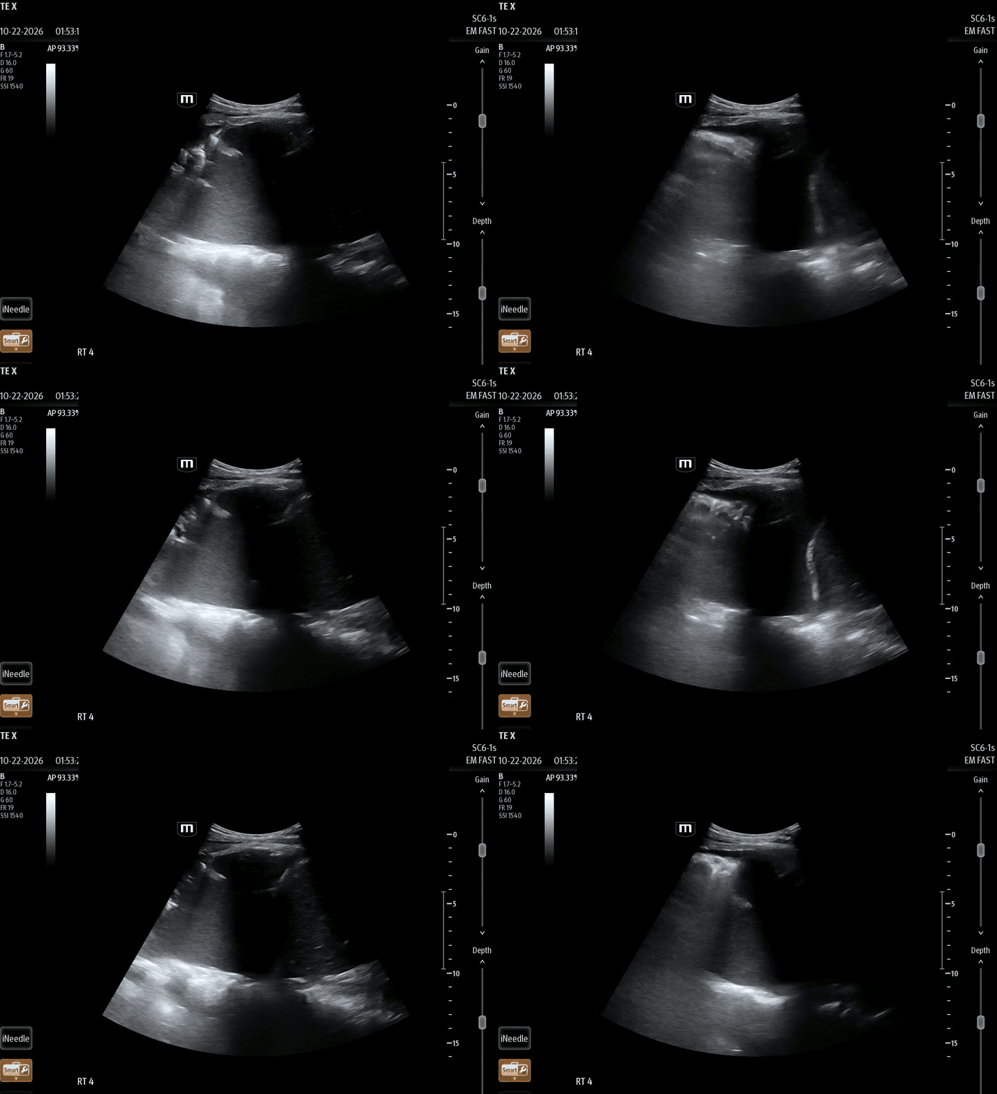
Figure 3: Spine sign
In pleural effusions, fluid is able to transmit ultrasound waves better than air so the spine becomes visible above the diaphragm - aka, spine sign.
Jellyfish Sign
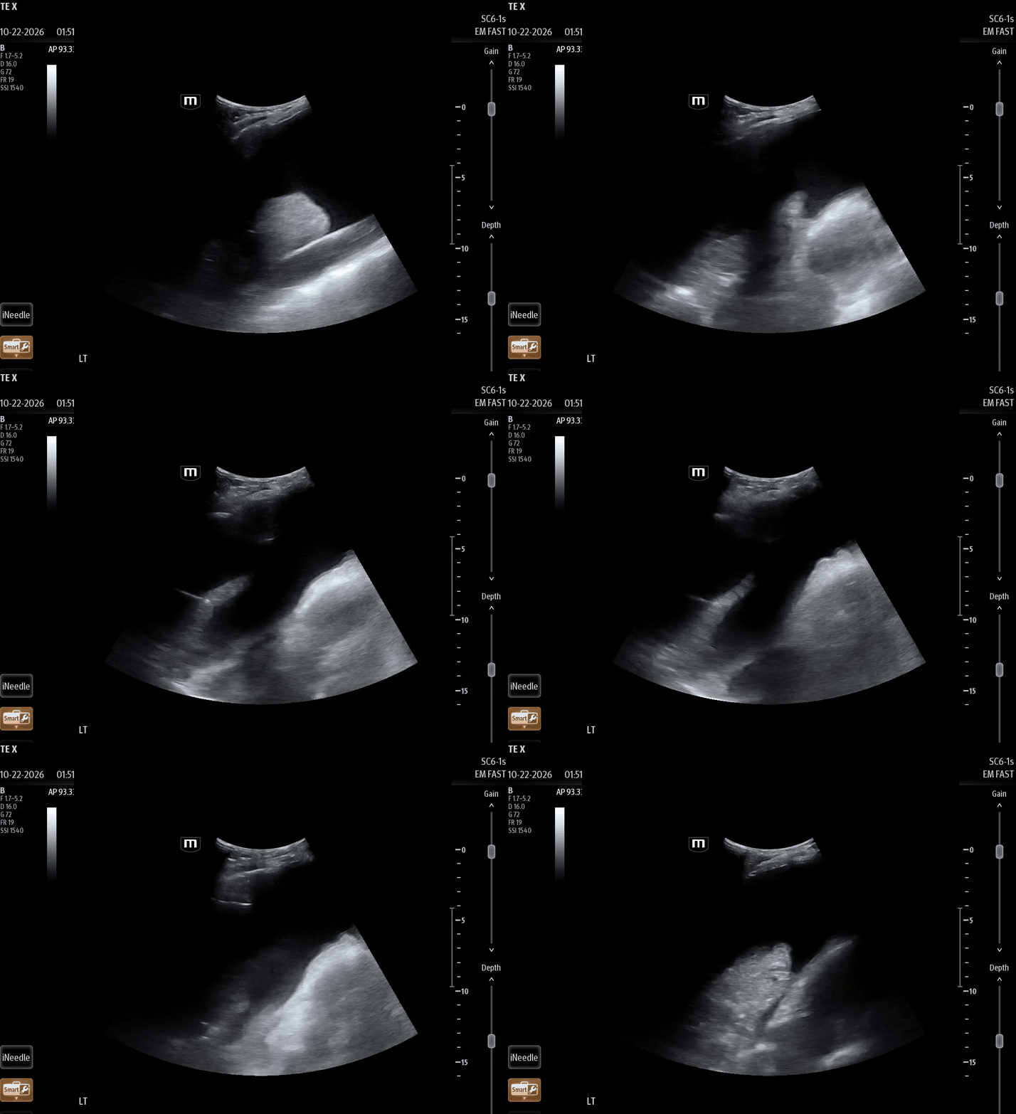
Figure 4: Jellyfish Sign
A compressed, atelectatic lung floats and moves within the anechoic effusion, resembling a jellyfish moving in water.
This has also been referred to as “lung tongue.”
Sinusoid Sign
When we apply M-mode to a normal lung, we get the Seashore sign (figure 5).
Above the pleural line (waves or sky) shows static, parallel horizontal lines representing stationary tissues of the chest wall.
Below the pleural line (beach) shows granular or sandy texture created by dynamic movement of the lung during respiration.
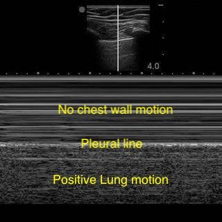
Figure 5: Seashore sign
When we apply M-mode to a suspected pleural effusion, we get the Sinusoid sign (figure 6).
The visceral pleura/lung line moves toward and away from the pleural line with respiration, producing a sinusoidal wave pattern.
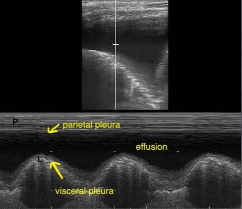
Figure 6: Sinusoid sign
Transudative or Exudative?
Transudative effusions have low protein and low cell count.
Ultrasound findings: simple anechoic fluid
Caution: this is not 100% specific to transudative effusions. Early exudative effusions can also appear anechoic.
Exudative effusions have high protein and high cell count, which causes echogenic particles to swirl within the fluid and look like plankton floating in the sea.
Plankton sign
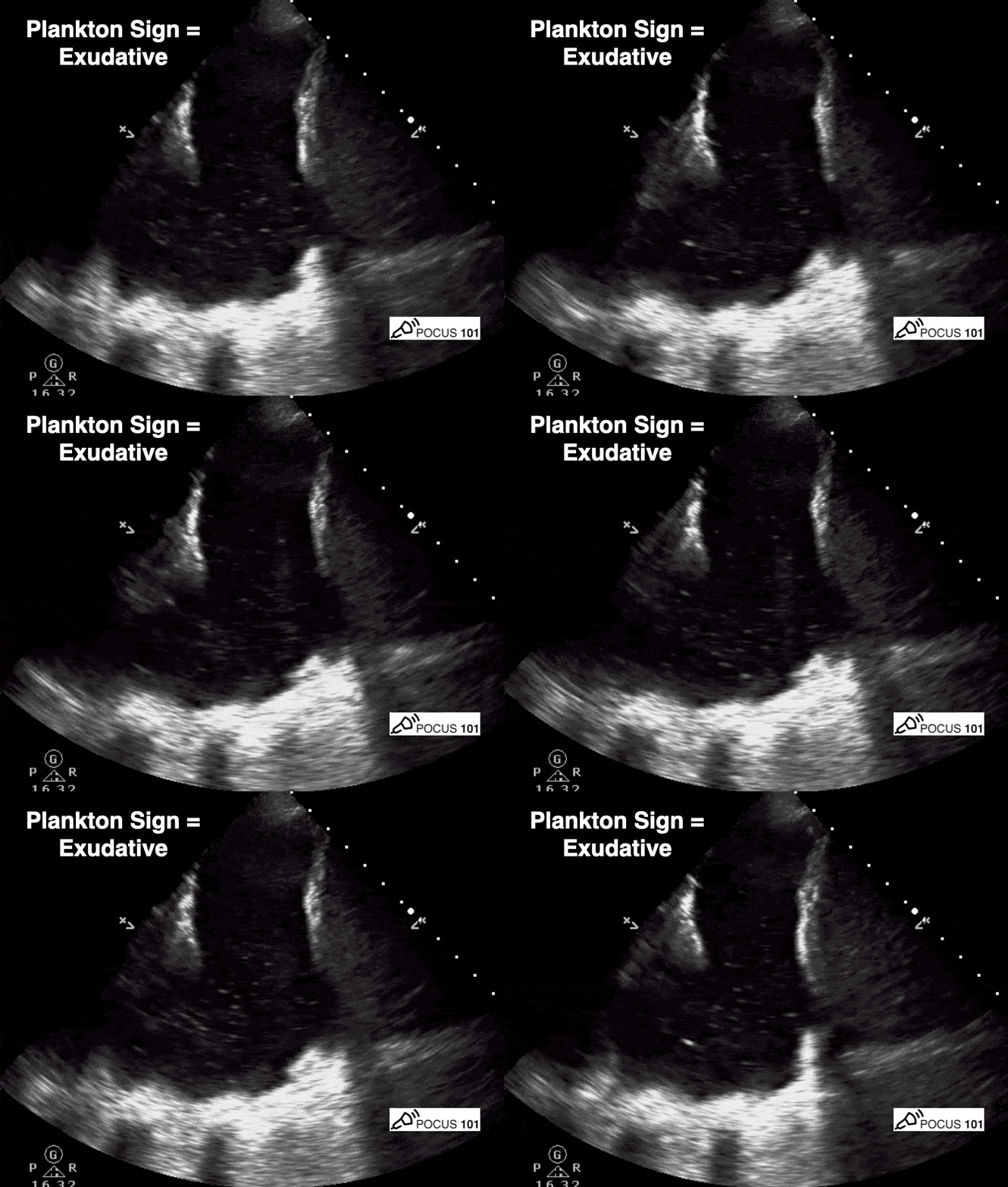
Figure 7: Plankton Sign
Septations/lobulations
Exudative effusions are primarily caused by processes like infection, inflammation, and malignancy that deposit fibrin into the fluid. As time passes, the fibrin strands organize into webs (septations) and eventually walled off pockets (loculations).
This has a reported 98% specificity and 94% positive predictive value for exudative fluid.
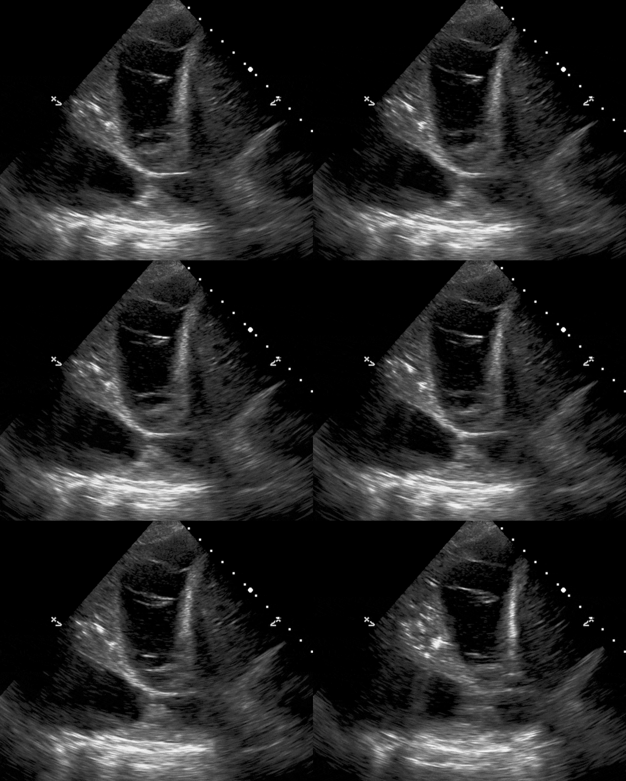
Figure 8: Septations/ Lobulations
Back to our case:
A left pigtail catheter was placed, with initial drainage of 1.5 L of serosanguinous fluid.
Cytology revealed adenocarcinoma.
VATS and talc pleurodesis were performed given the high likelihood of reaccumulation of malignant effusions.
Take home points:
Look for the spine sign and jellyfish sign to confirm a pleural effusion, and the sinusoid sign on M-mode.
Anechoic fluid suggests transudative fluid, but does not rule out exudative fluid.
Plankton sign and septations/lobulations strongly suggest an exudative effusion.
Always consider malignancy in a patient with a unilateral effusion and an unclear cause, even when the initial concern was something else (here, PE) — send off fluid analysis!
Happy scanning!
References
Bhoil R, Ahluwalia A, Chopra R, Surya M, Bhoil S. Signs and lines in lung ultrasound. J Ultrason. 2021;21(86):e225-e233.
Deschamps J, Dinh V, Ahn J, et al. Lung ultrasound made easy: step-by-step guide. POCUS 101. Accessed October 29, 2026. https://www.pocus101.com/lung-ultrasound-made-easy-step-by-step-guide/
Evans PT, Zhang RS, Cao Y, et al. The use of thoracic ultrasound to predict transudative and exudative pleural effusion. POCUS J. 2021;6(2).
Krishna R, Antoine MH, Alahmadi MH, Rudrappa M. Pleural effusion. In: StatPearls. StatPearls Publishing; 2024. Updated August 31, 2024. Accessed November 5, 2026. https://www.ncbi.nlm.nih.gov/books/NBK448189/