A Spooky Twist
Written by: Dr. Richard Gallardo
Edited by: Dr. Joann Hsu
Case Presentation
14 year old M with no significant PMH presenting for recurrence of left testicular pain and abdominal pain. Initially began suddenly yesterday at which time he presented to the ED and had an US negative for torsion. Pt tried taking Tylenol at home without relief. Also reports no known trauma to the testicles, dysuria, hematuria, nausea, or vomiting.
Diagnosis… Testicular Torsion!
Testicular Torsion Overview
Defined as twisting of the spermatic cord and is considered a SURGICAL EMERGENCY!
Accounts for about 10-15% of acute scrotal disease in children and has orchiectomy rates of about 42% in boys undergoing surgery
Of note, the presence of the cremasteric reflex does not confirm the absence of torsion
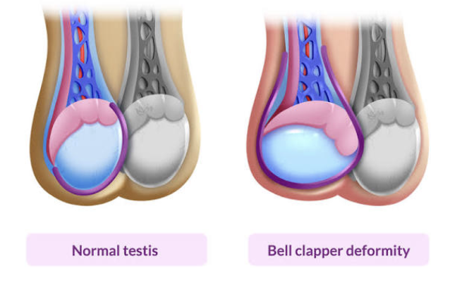
Presentation follows a bimodal distribution
First peak during the neonatal period - extravaginal torsion
May occur antenatally or in early postnatal period
Torsion of spermatic cord, epididymis, and testicle
Second peak is usually seen around puberty - intravaginal torsion
Most commonly associated with a bell clapper deformity - a congenital condition in which there is abnormal fixation of the testicle to the inner wall of the scrotum, thus, allowing for increased mobility of the testicle within the tunica vaginalis and predisposing patients to torsion
This predisposing anatomic condition is presumed to be bilateral and so patients must have both sides checked.
Symptoms
Sudden, severe pain in the scrotum
Scrotal swelling
Abdominal pain
Nausea
Vomiting
Testicular position that is high riding or unusual angle
Role of Ultrasound in Diagnosis
Sensitivity 88.9-100%; Specificity 97-98.8%
Linear probe
Provides great balance between penetration and resolution
Great for detecting low flow systems
Side-by-side comparison
Positioning
Place towel underneath scrotum for support
Drape the patient as modestly as possible, exposing only as much as needed to perform the ultrasound
Patient may require frog-leg position
Views
Buddy view
Short and long views, fanning completely through each of the views
Color doppler of each testicle to measure blood flow
Tips
Use enough gel to eliminate gas trapped in the skin folds
Do no change settings when moving from one testis to the other to keep the comparison accurate between the unaffected and affected testicle
Initial Imaging
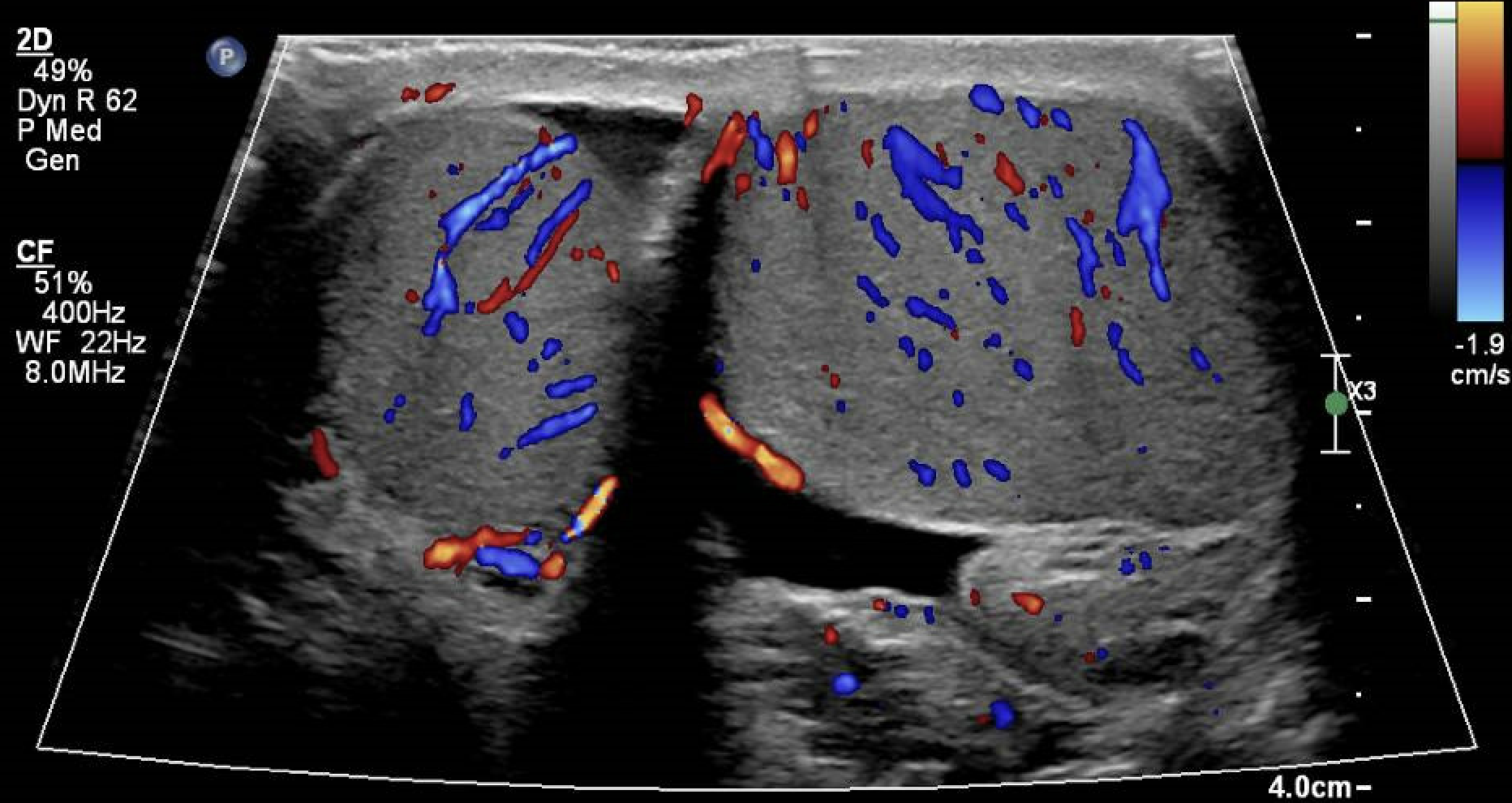
In this image from the patient’s initial visit, we note that there is adequate blood flow noted to bilateral testicles on gross visual assessment.
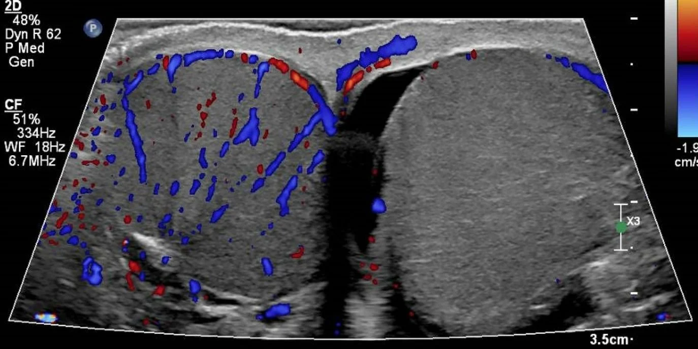
In this image from the patient’s second visit, we note that there is now no blood flow noted to the patient’s left testicles, highly concerning for torsion in this clinical context.
Short and Long Axis views
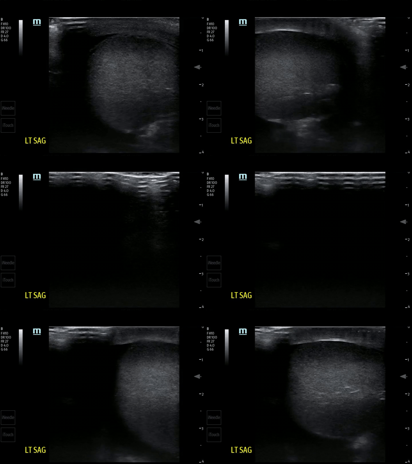
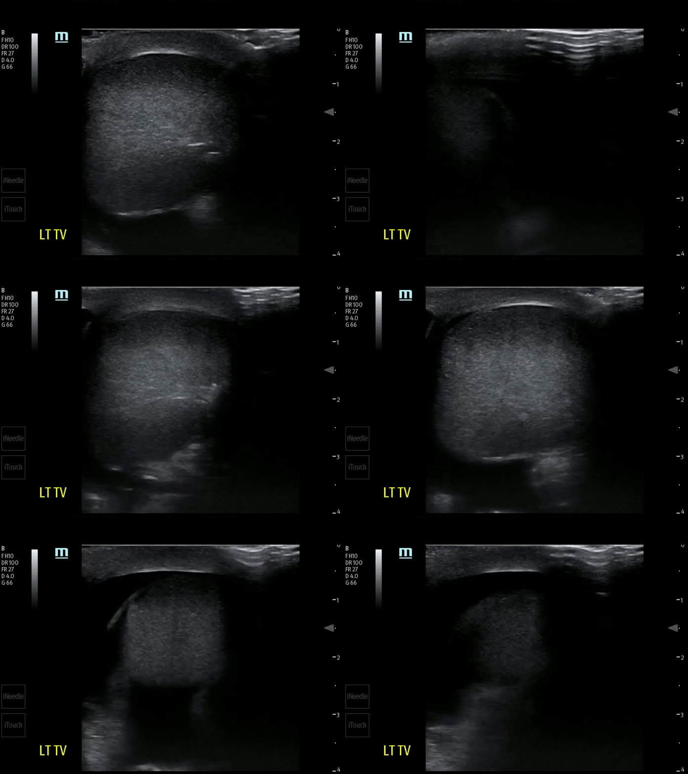
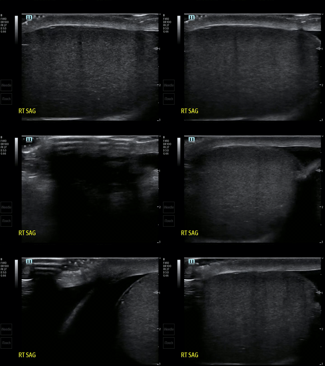
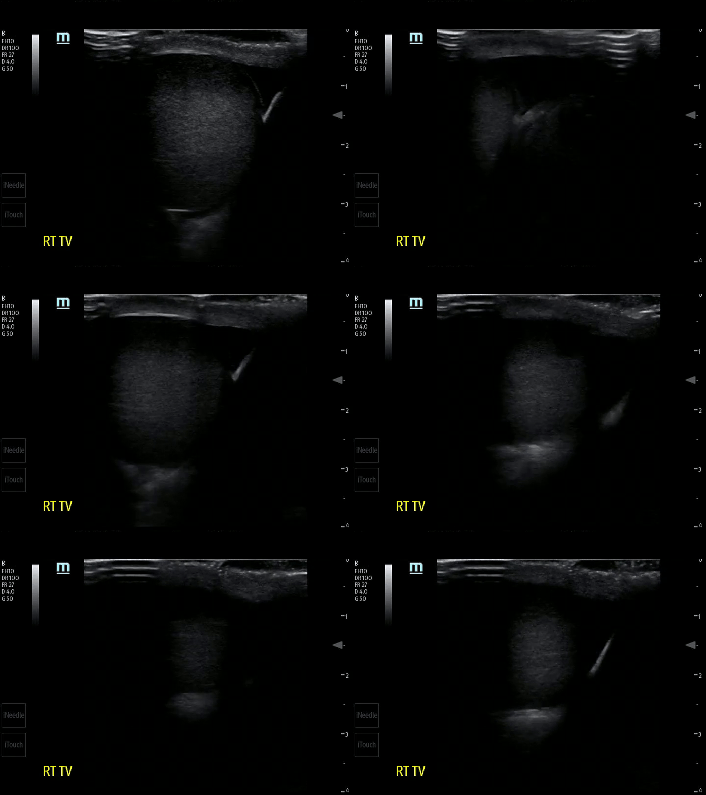
Testicular torsion is associated with many ultrasound findings. Below are some that can clue you in to a diagnosis of testicular torsion.
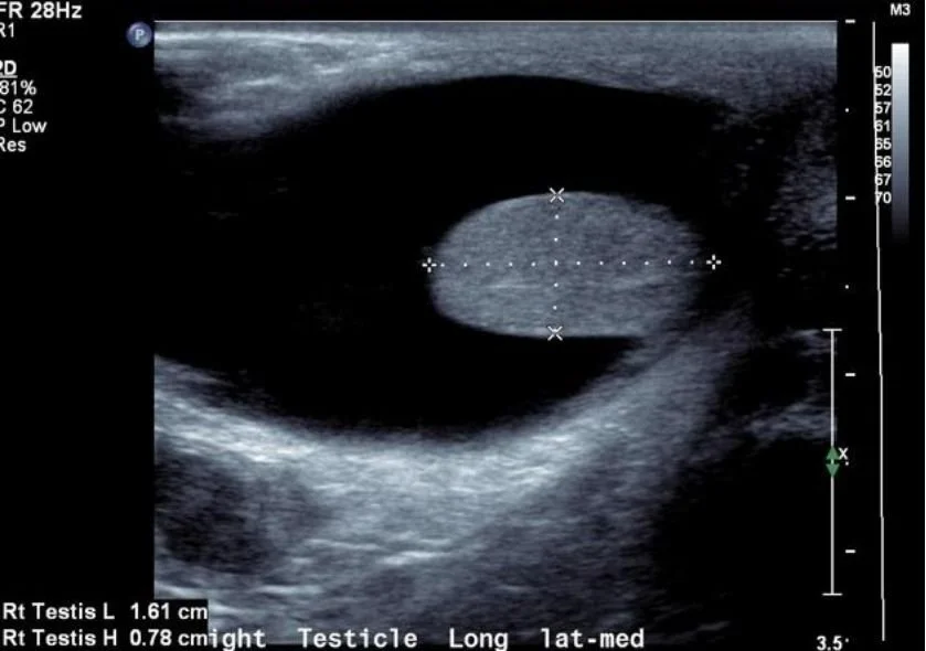
Reactive Hydrocele
Shown here is a hydrocele which is a finding that is sometimes associated with testicular torsion:
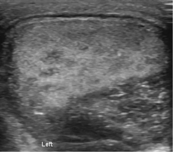
Heterogeneity
This is a testicles that appears to have heterogenous echotexture. This is a late finding (typically after 24 hours) and implies necrosis. The hypoechoic regions represent necrosis whereas the hyperechoic regions represent hemorrhage (can occur after reperfusion).
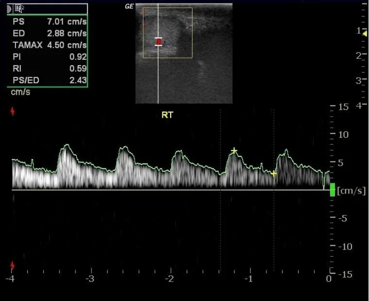
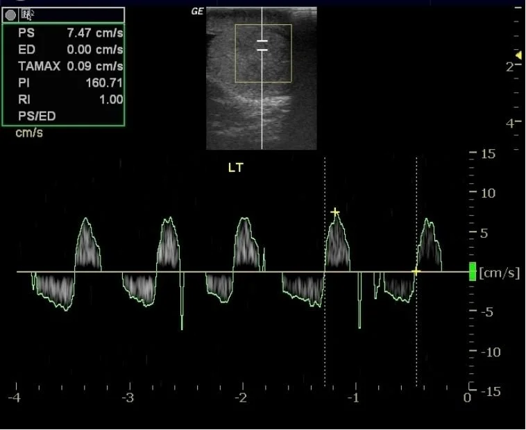
Resistive Index
Testicular torsion results in increased venous pressure and congestion with subsequent decreased arterial blood flow and ischemia. This translates to increased resistance of the vascular system and can be calculated as such. (Peak systolic flow - End diastolic flow) / Peak systolic flow. The image on the left represents a normal resistive index of <0.7. The image on the right represents a high resistive index of >0.7. In this right clinical context, this is suggestive of testicular torsion.
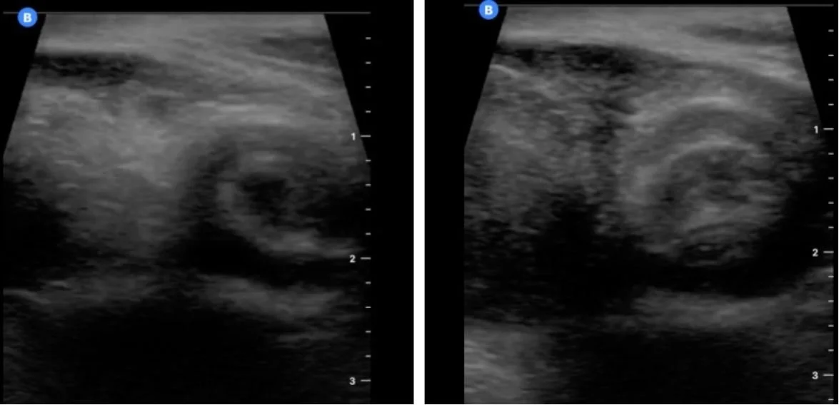
Whirlpool Sign
This shows what is called the “Whirlpool sign”. This is due to the twisting of the spermatic cord contents. It is the most specific and sensitive finding in both complete and incomplete torsion.
Treatment and Prognosis
Rapid diagnosis and treatment with surgical detorsion/orchiopexy
“Time is testicle”
<6 hours ~ 100% viability
6-12 hours ~ 50% viability
12-24 hours ~ 20% viability
Absent flow with heterogeneity suggests non-viability
Patient Follow-up
High concern for torsion based on bedside POCUS
Official US showed no blood flow in left testicle
Taken to OR for BL orchiopexy, both testicles viable
Discharged home same day
Happy scanning!
References
https://www.clinicalkey.com/#!/content/playContent/1-s2.0-S0002838X1360369X
https://www.mayoclinic.org/diseases-conditions/testicular-torsion/symptoms-causes/syc-20378270
https://pmc.ncbi.nlm.nih.gov/articles/PMC8112403/
https://www.sciencedirect.com/science/article/pii/S073386270400032X?via%3Dihub
https://radiopaedia.org/articles/testicular-torsion?lang=us