
Appendix ultrasound
Written by: Dr. Richa Gupta
Edited by: Dr. Joann Hsu
The case
4 year old comes in with lower abdominal pain, vomiting/diarrhea - what imaging do you want?
…ultrasound!
Let’s talk about RLQ ultrasound.
Probe Selection:
Pick between the linear and curvilinear probes for your patient - linear better for image quality, curvilinear better for depth. This will depend on the body habitus of the patient.
Landmarks:
Find the iliac crest and move medially. Identify your psoas muscle and iliac vessels. The appendix will typically course anteriorly to the psoas and iliac artery.

Here is a clip from our patient identifying the above landmarks:

You can see the psoas on the left and the iliac vessels just to the right of it.
Diagnosis:
Appendicitis is diagnosed based on the following criteria
Appendix AP measurement >6mm
Noncompressible
Tubular structure
Blind ending structure - aka not just a loop of bowel
Back to our patient:

This appendix diameter is within normal limits.
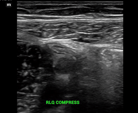
Here the appendix is compressible.
Additional Findings:
Not required for diagnosis but may be secondary signs of appendicitis.
Periappendiceal fat inflammation
Periappendiceal free fluid
Ring of fire
Appendicolith/fecolith

There is a fecolith here, but the appendix itself appears normal.
Takeaways/Tips
This patient did not have appendicitis! But finding a normal appendix on bedside ultrasound can often be more difficult than spotting appendicitis.
You may not see the appendix on bedside US, scans are specific not sensitive for appendicitis.
You will have the best luck at finding the appendix at the point of maximal tenderness as this is where an inflamed appendix, if present, is likely to be irritating the peritoneum.
For overlying bowel gas, use graded compression for better visualization.
You can try putting the patient in left lateral decubitus position to help identify the presence of a retrocecal appendix.
Happy scanning!
References