
Ocular ultrasound for elevated ICP
Written by: Dr. Kiran Kaur
Edited by: Dr. Joann Hsu
The case:
25-year-old male with no past medical history presented with worsening right eye vision changes for the past 10 months. He was in a physical altercation and experienced blunt-force trauma to the right eye.
Initially, he had episodic vision loss, however now he sees “black” constantly in his periphery. His central vision is intact. Denies any pain, headaches, or dizziness.
He was sent in by his ophthalmologist for an MRI.

Image 1 and 2: Normal Eye Anatomy vs the anatomy that can be visualized using ultrasound
ROLE OF ULTRASOUND
Ultrasound is indicated in patients with eye pain, acute vision changes, eye trauma, concern for increased intracranial pressure (ICP), or if there is too much surrounding periorbital edema to visualize the eye on external exam.
Optic nerve sheath diameter (ONSD) can be used to correlate with ICP elevation in a noninvasive way.
Idiopathic intracranial hypertension
Intracranial hypotension
Hypoxic-ischemic encephalopathy
Perioperative neurosurgical patients
Shunt evaluation
Head injury
Ischemic strokes- With every 0.1 cm increment in ONSD, the odds ratio for mortality increased 4.2 times.
Hematoma progression in hemorrhagic strokes
Systematic review and meta-analysis have shown that ONSD of more than 5.00 to 5.70 mm has a concurrent ICP value above 20 mm Hg
How to perform the ultrasound to measure optic nerve sheath diameter
Do NOT use ultrasound on the eye if there is any concern at all for globe rupture.
Use the high-frequency linear probe.
When doing an ultrasound of the eye you can place tegaderm as a barrier for the gel, however, you may see a-lines due to trapped air, which may impact scan quality.
You want to scan the eye in two planes: axial and sagittal; using fanning or sliding technique. Ask the patient to look left, right, up and down while in axial plane for a dynamic view.
Measure the optic nerve sheath diameter 3 mm distal to the posterior aspect of the globe, see below. Then, measure the diameter of the entire optic nerve sheath across. Normal diameter is less than 5 mm.

Image 3: Ultrasound of the eye with measurements for optic nerve sheath diameter.
Back to the case:
Here are the POCUS images for this patient.

Image 4: Left eye sheath diameter measurement

Image 5: Right eye sheath diameter measurement
The right eye had optic disc elevation with bilateral dilated optic sheath concerning for increased intracranial pressure. The left eye diameter was measured to be 69 mm and the right eye was 70 mm.
Ocular pathology seen on ultrasound
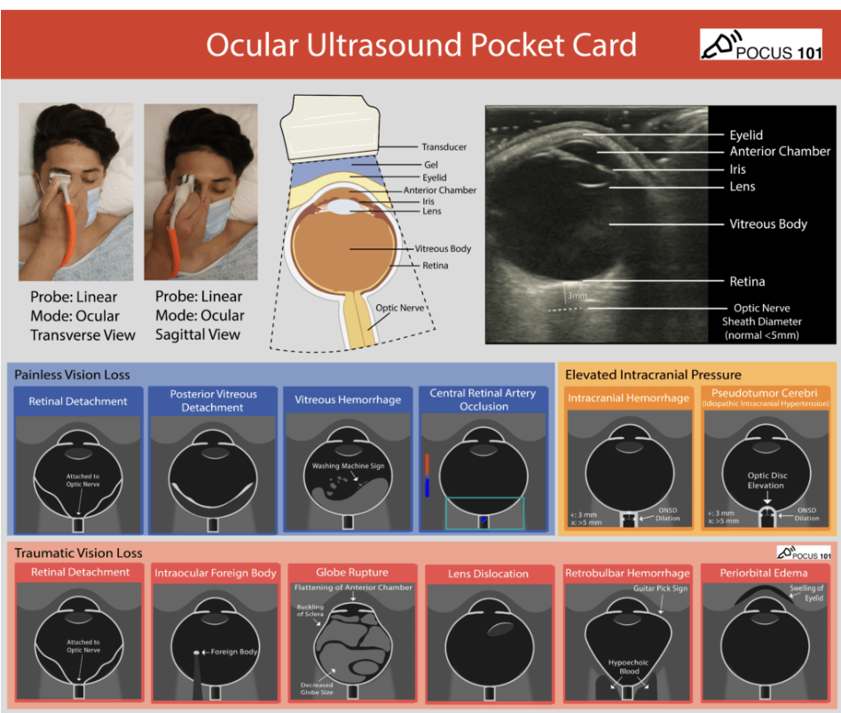
Ultrasound is useful in diagnosing many pathologies, some of which are emergencies and require emergent ophthalmology consults. If there is suspicion for globe rupture, end the scanning immediately.
END OF THE CASE
The patient had a MR Orbit and Brain with and without contrast, which was unremarkable, however there was partially empty sella, which can be seen in idiopathic intracranial hypertension MR Angiography of Head and Neck was unremarkable.
MR Venography was concerning for non-specific enhancement, which was follow up with CT Venography of the brain, and the patient was found to have Venous Sinus Thrombus
The patient was started on Acetazolamide. A week later, he was seen in our ER again, and his vision had improved.
Happy scanning!
References
https://www.pocus101.com/ocular-ultrasound-made-easy-step-by-step-guide/
https://www.ncbi.nlm.nih.gov/books/NBK554479/#:~:text=Point%20of%20care%20optic%20nerve,value%20above%2020%20mm%20Hg.