
SCPB - The Neck’st best thing!
Written by: Dr. Justin Song
Edited by: Dr. Joann Hsu
Case
61 year-old male with PMHx of HTN, HLD, MI, CAD s/p stent (2019), DM2, UC, on ASA, presenting after MVA. Ejected from his scooter without a helmet after hitting a pothole while driving approximately 20mph. He endorses head strike, but he is unsure of LOC. Complains of pain mostly to his right chest where he landed. Denies any vision changes, N/V, fevers, abdominal pain, N/T to extremities.
Pertinent exam:
CHEST: (+) pain and TTP R lateral ribs; (-) crepitus
EXTREMITY: (+) pain and TTP of anterior lateral R shoulder, (+) R 5th digit dislocation, (+) full ROM intact upper and lower extremities otherwise. (+) strong DP pulses
Imaging: CT: fractures of the right 1st-7th ribs; and fracture of the right clavicle (at the medial margin of a fixation plate).

Disposition
Admitted to trauma team for further evaluation and management.
Consulted ortho who recommends NWB RUE and will follow inpatient course
Patient agreed to ESPB and SCPB as part of multimodal pain management offered by US team
What is SCPB?
Superior cervical plexus block (SCPB) is a straightforward regional anesthesia technique for interventions involving C2-C4 sensory nerves
Dermatomal distribution along the anterolateral neck, anterior and retro-auricular areas, submandibular area, cape region along the shoulder and clavicle
Procedures include carotid endarterectomy, thyroid/parathyroidectomy, and LN excision. In the ED, this nerve block is often used for IJ CVC placement or abscess drainage/laceration repairs i
Possible complications involve local infections, hematoma formation, local anesthetic systemic toxicity (LAST).
Its counterpart, the deep cervical plexus block (DCPB) carries a higher risk for phrenic nerve block (diaphragmatic paresis), and inadvertent subarachnoid/epidural anesthesia
Anatomy
The superficial cervical plexus (SCP) originates from the C2-C4 nerve roots and is located deep to the sternocleidomastoid muscle (SCM), and superficial to the prevertebral fascia that contains the levator scapulae (LS) and scalene muscles
Four terminal branches emerge from the midpoint of the posterior border of the SCM at approximately the level of the thyroid cartilage: lesser occipital nerve, greater auricular nerve, transverse cervical nerve, and supraclavicular nerves

Protocol and Materials
The materials used in the SCPB block are the same as any other nerve block, but one exception is that a standard 25-gauge needle may be used instead of a long spinal needle given the superficial location of the nerve bundle.
Important points to note from the list include the use of Ropivacaine without epinephrine as the choice of analgesic because it has reduced cardiotoxicity and longer duration of action compared to its long-acting counterpart bupivacaine and it’s shorter acting alternative, lidocaine.
Also, patients will need to be placed on telemetry and require lipid emulsion therapy at bedside in case of systemic anesthetic toxicity
Technique
Patient is supine and has their head rotated contralateral to the side of injury with the operator in the ipsilateral side.
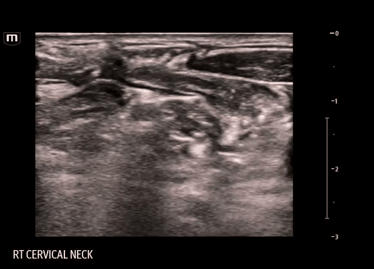
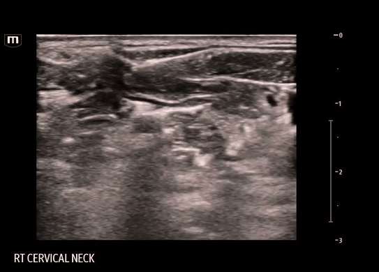
Using a linear probe, start scanning over the SCM at the level of the thyroid cartilage and slide laterally until visualization of the edge of the sternocleidomastoid muscle (SCM), underlying levator scapulae (LS) muscle, internal jugular vein (IJV) and carotid artery (CA).
SCP nerve bundle may or may not be present below the SCM
This procedure is done with in-line technique and the target is the fascial plane just deep to the SCM and superficial to the prevertebral fascia containing the LSM and scalene muscles.
Enter on the opposite side of the SCM so the tip of your needle encounters the tapered end of the SCM
Be careful as the external jugular vein (EJV) lies in close proximity to the edge of SCM
Apply constant negative pressure aspiration until you reach the target
You may trial small amounts of saline to hydrodissect the plane and confirm the correct location
Deposit 10-15ml of local anesthetic into the space
The SCPB does not require a large amount of anesthetic to be effective
Always calculate the max dose of anesthetic for your patient based on weight, choice of anesthetic, and comorbidities. The app “safe local” is a great resource for this.
It is important to visualize the needle tip during the procedure.
Remember the probe is already in the ideal position with all the landmarks identified, so the needle will have to be corrected with micromovements to stay in the window of the probe.
In a successful SCPB, you will see the SCM lift off from the prevertebral fascia


Clips from our block:
FYI: During this particular block, the needle entered from the same side as the SCM, contrary to the usual technique. This was done to avoid a vessel on the other side. The block should routinely be done as described above.
Outcome
ESPB was successful since patient reported improvement in his rib pain. However, patient continued to endorse pain over his right clavicle, which suggests the SCPB was not successful.
In retrospect, there are several reasons why the SCPB may not have worked for our patient:
Insufficient analgesia:
Literature review shows surgical repair of clavicular fracture can be done with regional anesthesia with a combination of interscalene and cervical plexus block since the clavicle is co-innervated by the branches of the cervical and brachial plexus. SCPB alone may not have been sufficient enough for our patient
Poor technique:
For our patient, we decided to approach the facial plane from medial-to-lateral through the SCM instead of the lateral-to-medial towards the edge of the SCM due to the close proximity of the EJV. Approaching the fascial plane perpendicularly appears to have diverted our target trajectory and the anesthetic was consequently injected into the LS.
Happy scanning!
References
Dr Bagalkot says: (n.d.). Ultrasound-Guided interscalene brachial plexus nerve block. Core EM. https://coreem.net/core/ultrasound-guided-interscalene-brachial-plexus-nerve-block/
Hipskind, J. E., Hendrix, J. M., & Ahmed, A. A. (2024, March 2). Cervical plexus block. StatPearls - NCBI Bookshelf. https://www.ncbi.nlm.nih.gov/books/NBK557382/
Operater. (2024, November 1). Ultrasound-Guided cervical plexus nerve block. NYSORA. https://www.nysora.com/techniques/head-and-neck-blocks/cervical/ultrasound-guided-cervical-plexus-block/
Pain control using Ultrasound-Guided superficial cervical plexus block. (2015, June 8). ACEP Now. https://www.acepnow.com/article/pain-control-using-ultrasound-guided-superficial-cervical-plexus-block/
Regional Anesthesiology and Acute Pain Medicine. (2020, October 7). Interscalene Brachial Plexus block [Video]. YouTube. https://www.youtube.com/watch?v=5_EKD83UvNY
Regional Anesthesiology and Acute Pain Medicine. (2021, October 17). Superficial cervical plexus block [Video]. YouTube. https://www.youtube.com/watch?v=hmfvUldlvfY
Rose, G., & Pumarejo, L. (2022, September 12). Ultrasound-Guided Superficial Cervical Plexus Block. ACEP. Retrieved October 26, 2025, from https://www.acep.org/emultrasound/newsroom/september-2022/ultrasound-guided-superficial-cervical-plexus-block