
Unmasking Sepsis with POCUS
Written by: Dr. Khin Oo
Edited by: Dr. Joann Hsu
Case:
55 year old male with no PMHx presenting to ED with complaints of rectal pain, left lower extremity swelling, chest pain. Patient is a poor historian, but A&Ox3. States that over the last 5 days, he has noticed worsening left lower extremity swelling associated with chest pain, difficulty breathing, cough. He denies known fevers, palpitations, dizziness, congestion, history of previous symptoms, recent travel.
Vitals:
BP 113870 | HR 130 | RR 16 | T 36.4 C | SpO2 96% RA
Pertinent exam:
EXTREMITY: Left lower leg (+) edema 2+, (+) crepitus, (+) erythema (+) full ROM intact upper and lower extremities otherwise. (+) DP pulses
Using the linear probe for better visualization of superficial structure, let’s take a look at what we saw on the left lower leg:
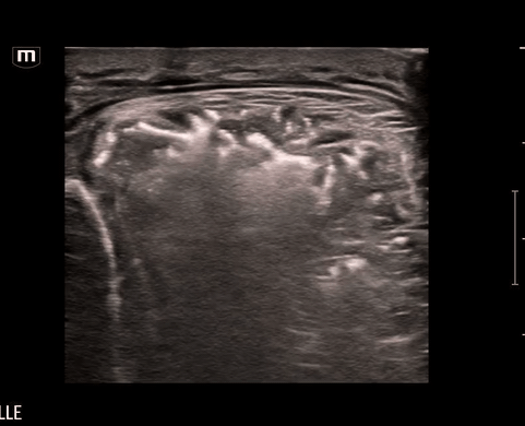
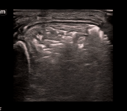
What do we see?? Air and “dirty shadowing” in the soft tissue concerning for necrotizing fasciitis in this clinical context.
We can identify necrotizing fasciitis on US by using the acronym “STAFF”
S – Subcutaneous thickening: diffuse thickening and distortment of soft tissue layers seen
T – Thickened fascia: chunky fascia not a thin bright white line
A – Air: posterior dirty acoustic shadowing, corresponding to gas in the soft tissue
F – Fascial fluid: Anechoic fluid collections (<2mm)
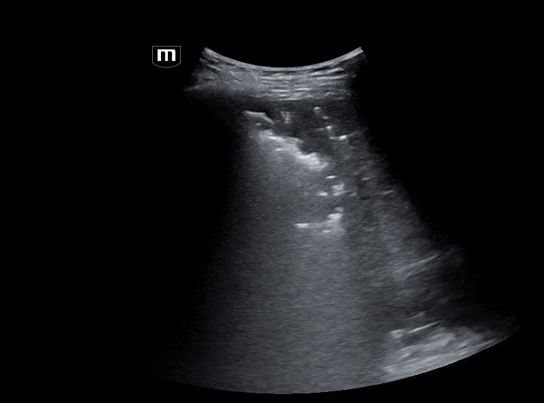
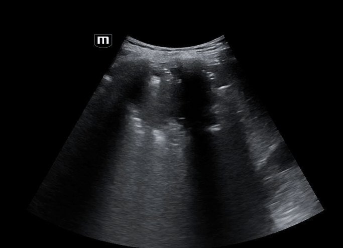
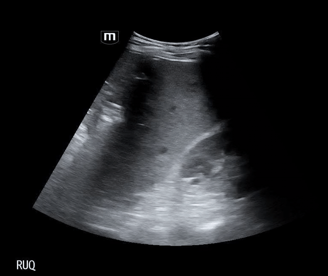
The patient was a limited historian and reported multiple complaints which included chest pain, left lower extremity pain, rectal pain, and cough. Due to the unclear history and concern for potential underlying pathology, a bedside FAST ultrasound was obtained.
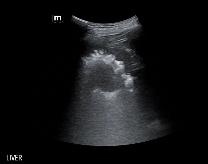
Using the curvilinear probe, we see a well-defined, round lesion within the liver parenchyma on the right lower quadrant US:
Let’s talk about some differential diagnoses:

Based on the patient’s ultrasound findings, the appearance is consistent with a hepatic abscess.
Hepatic Abscess: Key Sonographic Features
Appearance: focal, hypoechoic or complex cystic lesion within liver parenchyma
Echotexture: heterogeneous, may contain septations, debris, or internal echoes
Borders: irregular or ill-defined; chronic abscesses may develop capsule
Gas formation: echogenic foci with dirty shadowing or ring-down artifacts (seen in Klebsiella/E. coli)
Posterior acoustic enhancement: fluid-filled lesion characteristic
Color Doppler: peripheral hyperemia, absent central vascularity
Location: most commonly in the right hepatic lobe due to its greater blood supply and biliary drainage.
What are the different types of hepatic abscess and management?

Back to the Case
Significant Labs
Metabolic Panel:
Sodium: 124 mmol/L (Hyponatremia)
Calcium: 7.0 mg/dL (Hypocalcemia)
Bicarbonate: 15 mmol/L (Low)
Anion Gap: 18 (Elevated)
Glucose: 327 mg/dL (Hyperglycemia)
Hemoglobin A1C: 12.4% (Poor long-term glycemic control)
Liver Function Tests:
Total Bilirubin: 1.5 mg/dL
Direct Bilirubin: 1.0 mg/dL (Mild hyperbilirubinemia)
AST: 384 U/L
ALT: 339 U/L (Transaminitis)
Alkaline Phosphatase: 335 U/L (Elevated)
Inflammatory Markers:
C-Reactive Protein (CRP): 19.6 mg/L (Elevated)
Lactic Acid: 2.8 mmol/L (Lactic Acidemia)
Diagnosed with Disseminated Klebsiella pneumoniae infection (hypervirulent strain) with the following complications:
Necrotizing intramuscular and soft tissue infection with abscess formation within the anterior tibialis muscle, extending to the mid-femoral diaphysis and posterior distal thigh
Septic emboli with associated pulmonary infarcts
Multiple hepatic abscesses, the largest located in the right hepatic lobe, measuring 13.3 cm
Acute prostatitis
Right temporal lobe brain abscess
Management
Meropenem and linezolid for broad-spectrum IV antibiotics
IR percutaneous drainage of hepatic abscess
OR debridement and washout of left lower extremity necrotizing fasciitis:
Take Home Points
STAFF acronym helps identify NF on ultrasound
Abscess = complex, irregular, debris-filled lesion
Clinical exam + POCUS = faster, smarter diagnosis
POCUS can rapidly identify infection sources and guide management in an undifferentiated septic patients
Happy scanning!
References
Yen, Z.-S., Wang, H.-P., Ma, H.-M., Chen, S.-C. and Chen, W.-J. (2002), Ultrasonographic Screening of Clinically-suspected Necrotizing Fasciitis. Academic Emergency Medicine, 9: 1448-1451. Lorem ipsum dolor sit amet, consectetur adipiscing elit. Vivamus quis ultrices felis. Fusce sapien nunc, posuere at mauris sed, sagittis luctus erat. Integer sollicitudin pellentesque dolor ac suscipit. Duis quis commodo mauris.
Magalhães, L., Martins, S.R.P. & Nogué, R. The role of point-of-care ultrasound in the diagnosis and management of necrotizing soft tissue infections. Ultrasound J 12, 3 (2020). https://doi.org/10.1186/s13089-020-0153-4
Marks A, Patel D, Sundaram T, Johnson J, Gottlieb M. Ultrasound for the diagnosis of necrotizing fasciitis: A systematic review of the literature. Am J Emerg Med. 2023 Mar;65:31-35. doi: 10.1016/j.ajem.2022.12.037. Epub 2022 Dec 22. PMID: 36580698.
Wang WJ, Tao Z, Wu HL. Etiology and clinical manifestations of bacterial liver abscess: A study of 102 cases. Medicine (Baltimore). 2018 Sep;97(38):e12326. doi: 10.1097/MD.0000000000012326. PMID: 30235686; PMCID: PMC6160130.
Lin AC, Yeh DY, Hsu YH, Wu CC, Chang H, Jang TN, Huang CH. Diagnosis of pyogenic liver abscess by abdominal ultrasonography in the emergency department. Emerg Med J. 2009 Apr;26(4):273-5. doi: 10.1136/emj.2007.049254. PMID: 19307388.
Buttar S, Cooper D Jr, Olivieri P, Barca M, Drake AB, Ku M, Rose G, Siadecki SD, Saul T. Air and its Sonographic Appearance: Understanding the Artifacts. J Emerg Med. 2017 Aug;53(2):241-247. doi: 10.1016/j.jemermed.2017.01.054. Epub 2017 Mar 31. PMID: 28372830.