Spondylo-WHAT?
Written by: June Chu, MD, MPH; Edited by: Timothy Khowong, MD, MSEd
Understanding the Terminology
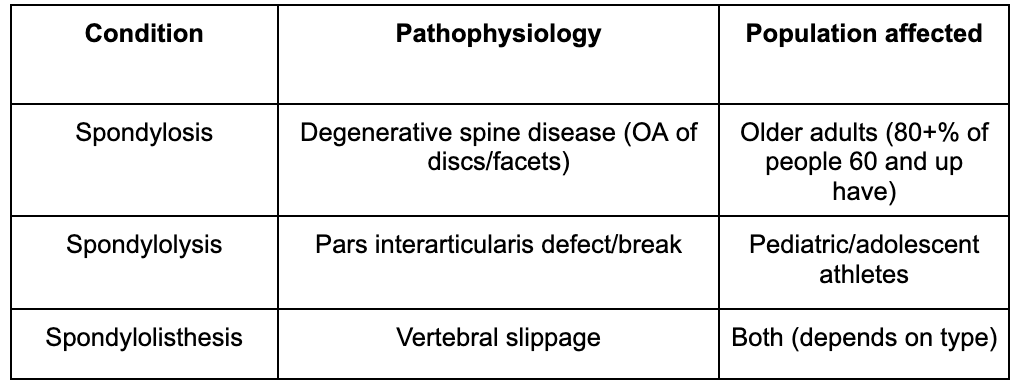
Meet the Spontaneous Spondylo Siblings: Spondylosis, Spondylolysis, and Spondylolisthesis
Derived from Greek where:
Spondylo = vertebra
-osis = process/degeneration
-lysis = break
-listhesis = slippage
Background
Back pain accounts for millions of ED visits annually. While most patients have benign mechanical pain, emergency physicians must rapidly distinguish degenerative disease from unstable spinal pathology, spinal cord compression, cauda equina syndrome, infection, malignancy, or traumatic injury.
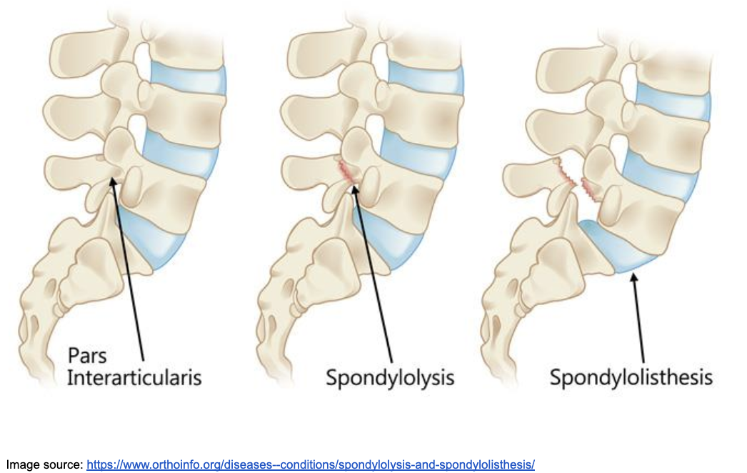
Spondylosis refers to degenerative osteoarthritis of the spine. Spondylolysis is a defect or stress fracture of the pars interarticularis, most commonly involving L5. Spondylolisthesis is translation of one vertebral body relative to another and may result from pars defects, degeneration, trauma, or congenital abnormalities.
Spondylosis
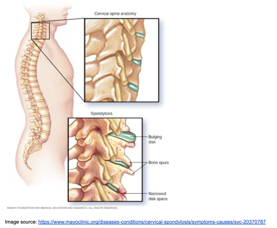
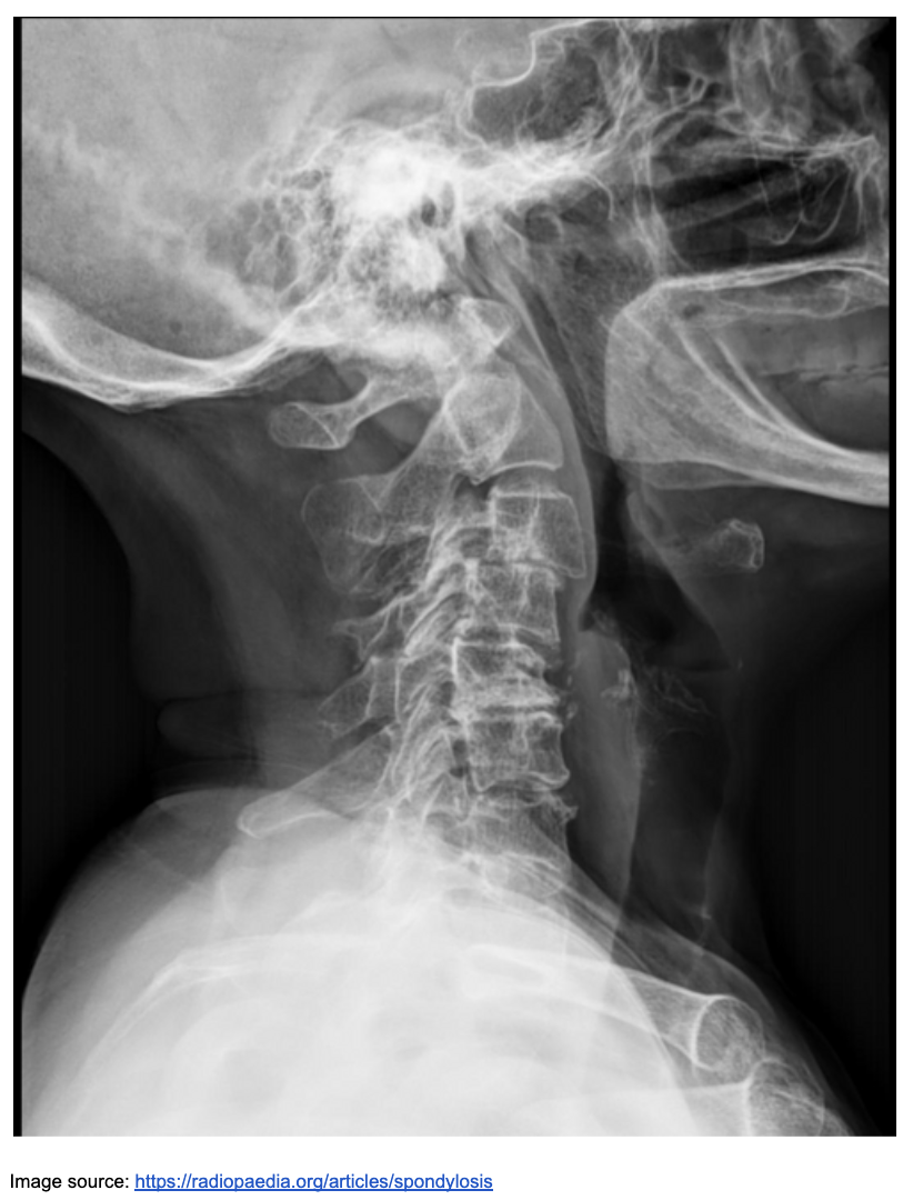
Spondylosis primarily affects older adults, with a higher prevalence in men than women. Common risk factors include occupations involving heavy lifting or professional driving, smoking, and participation in certain sports. The condition arises from intervertebral disc degeneration due to desiccation and loss of disc height, facet joint arthritis or hypertrophy, osteophyte formation, and various ligamentous changes.
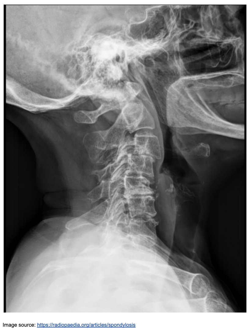
Potential complications include the narrowing of foramina leading to radiculopathy and central stenosis, which can result in myelopathy. Imaging typically begins with X-rays to identify osteophytes and disc space narrowing. While CT scans provide excellent bony detail, MRI is the most useful modality in the emergency department for evaluating neural compression in patients with neurological deficits.
Treatment generally focuses on conservative management with NSAIDs and muscle relaxants. Acute intervention is typically reserved for cases involving significant neurological deficits or suspected spinal cord compression. While the prognosis is generally chronic and progressive, spondylosis is frequently an incidental finding on imaging.
Spondylolysis
Spondylolysis is a very common condition, affecting up to 6-7% of adolescent athletes and implicated in up to 47% of low back pain complaints in this population. It typically involves a defect in the pars interarticularis of the L5 vertebra, which can progress to spondylolisthesis—the anterolisthesis of L5 relative to S1.
Risk factors are particularly high for athletes such as gymnasts, weight lifters, and football linebackers who engage in repetitive hyperextension. While many cases are asymptomatic and there is no direct association between radiologic grade and clinical presentation, a healthy active adolescent may present with acute onset of low back pain during athletic activity.
Imaging techniques vary, with oblique X-rays showing the classic “Scottie dog with collar,” CT providing the best view of bony defects, and MRI or SPECT being useful for detecting early stress reactions before a fracture occurs.
Treatment is primarily conservative, focusing on activity restriction and physical therapy. The prognosis is generally positive, as the majority of patients improve and early lesions have the potential to heal, although a small subset may progress to vertebral slippage.
Spondylolisthesis
The pathophysiology of spondylolisthesis involves the forward (anterolisthesis) or backward displacement of one vertebral body relative to another. This condition can be caused by isthmic factors such as a pars defect from spondylolysis, degenerative changes in the discs and facets, or congenital and traumatic origins. Imaging evaluation typically begins with lateral X-rays to identify vertebral slippage, while flexion-extension views are utilized to assess for instability. Additionally, MRI is used to evaluate for nerve compression, and CT scans provide detailed views of any underlying pars defects.
Management strategies are divided into non-operative and operative approaches. Non-operative management may involve observation alone for asymptomatic patients, regardless of slip grade, as they typically remain asymptomatic, though return to contact sports remains controversial. Physical therapy and activity restriction for six months—including hamstring stretching, pelvic tilts, and abdominal strengthening—are indicated for symptomatic spondylolysis and low-grade spondylolisthesis, with most patients improving without surgery. Acute pars stress reactions or cases failing physical therapy may require bracing, which has been shown to be superior to activity restriction alone. Operative management is reserved for those who fail conservative treatment. Pars interarticularis repair is generally preferred over fusion as it preserves motion; however, fusion may be necessary for patients with neurological deficits, provided that overaggressive reduction is avoided to prevent further impairment.
Evaluation:
History should identify trauma, fever, cancer, immunosuppression, IV drug use, anticoagulation, bowel or bladder dysfunction, saddle anesthesia, progressive weakness, gait changes, and duration of symptoms. Examination should include complete motor, sensory, reflex, gait, rectal tone when indicated, and assessment for upper motor neuron findings.
Imaging Pearls:
Avoid routine MRI for uncomplicated mechanical back pain. Obtain MRI urgently for suspected cord compression, cauda equina syndrome, epidural abscess, or severe neurologic deficits. CT is preferred for detailed bony anatomy, especially pars defects.
Red Flags:
Immediate spine consultation and MRI are indicated for bowel/bladder dysfunction, saddle anesthesia, progressive motor weakness, hyperreflexia with myelopathy, severe bilateral radiculopathy, or suspected spinal cord compression.
Disposition:
Discharge patients with reassuring examinations, controlled pain, and reliable follow-up. Admit or obtain emergent consultation for neurologic compromise, inability to ambulate, unstable traumatic injury, or concern for infection or malignancy.
Clinical Pearls:
Do not anchor on incidental degenerative findings. Imaging abnormalities often do not correlate with symptoms. Always correlate radiographic findings with a careful neurologic examination. Missing evolving cord compression has far greater consequences than overcalling degenerative disease.
References:
https://www.orthoinfo.org/diseases--conditions/spondylolysis-and-spondylolisthesis/
https://pmc.ncbi.nlm.nih.gov/articles/PMC9425520/
https://www.orthobullets.com/spine/2029/cervical-spondylosis