Joint Venture: Elbow Dislocation Edition
Written By: Khin Oo, MD; Edited by: Timothy Khowong, MD, MSEd
Elbow Anatomy: Osseous & Soft tissue:
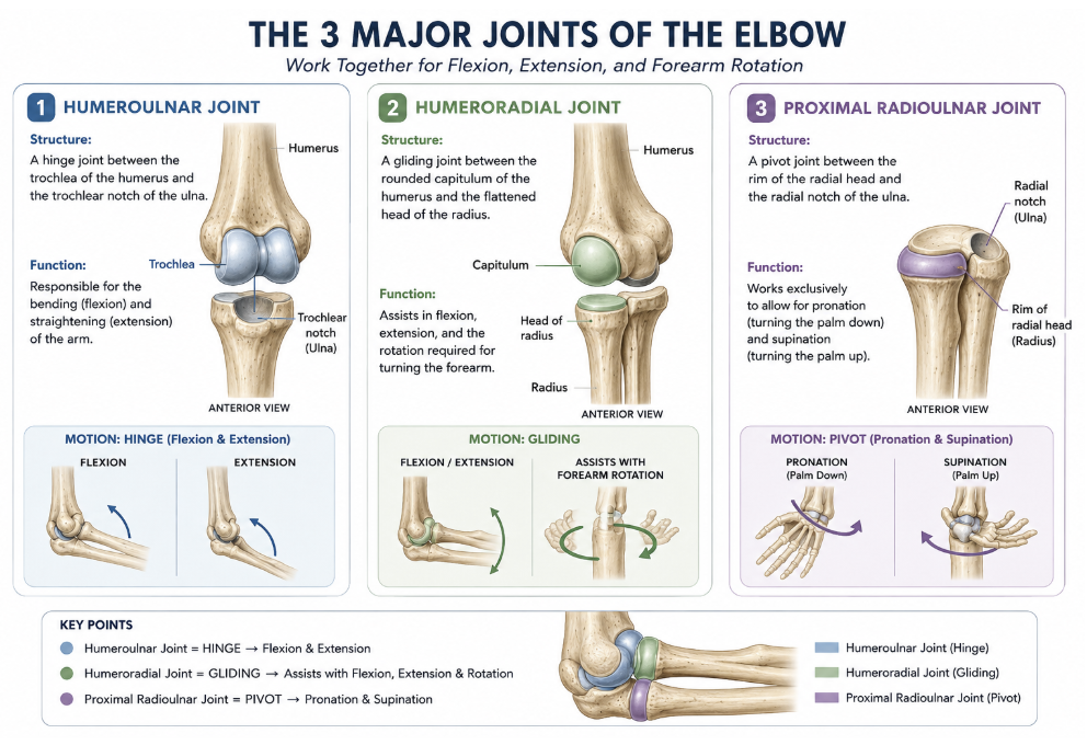
Osseous Stabilizers: the elbow is a trochoginglymoid (hinged-pivot) joint composed of three articulations:
1. Humeroulnar Joint Structure: A hinge joint between the trochlea of the humerus and the trochlear notch of the ulna. Responsible for the bending (flexion) and straightening (extension) of the arm
2. Humeroradial Joint Structure: A gliding joint between the rounded capitulum of the humerus and the flattened head of the radius. Assists in flexion, extension, and the rotation required for turning the forearm.
3. Proximal Radioulnar JointStructure: A pivot joint between the rim of the radial head and the radial notch of the ulna. Works exclusively to allow for pronation (turning the palm down) and supination (turning the palm up)
Soft Tissue Stabilizers
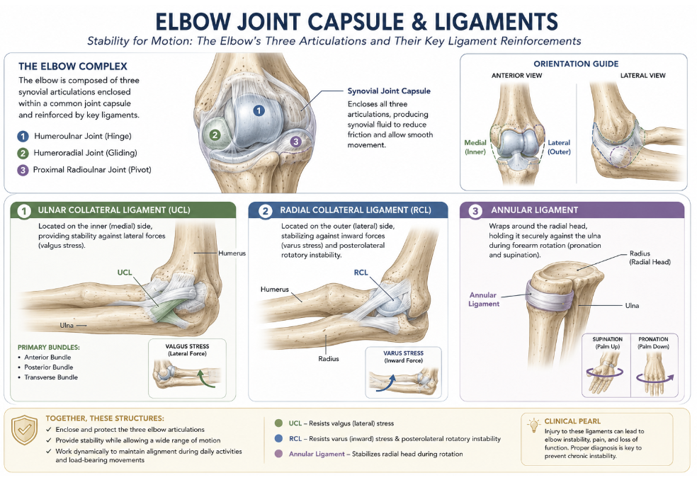
Lateral collateral ligament complex (LCL) - most important component is the lateral ulnar collateral ligament (LUCL), which resists posterolateral rotatory instability
Medial collateral ligament complex (MCL) - the anterior bundle is the primary restraint to valgus stress; most important soft tissue stabilizer of the elbow
Annular Ligament - Wraps around the radial head, holding it securely against the ulna during forearm rotation
Mechanism of Injury
Most common: fall onto an outstretched hand (FOOSH)
92% occurred at or near full extension, with a valgus moment being the most common deforming force
Clinical Evaluation
History & Physical Examination
Mechanism (FOOSH, direct blow, sports injury)
Dominant hand, occupation, sport
Prior elbow injury or surgery
Inspection: Obvious deformity; olecranon prominence posteriorly; swelling
Neurovascular assessment (Before AND After reduction):
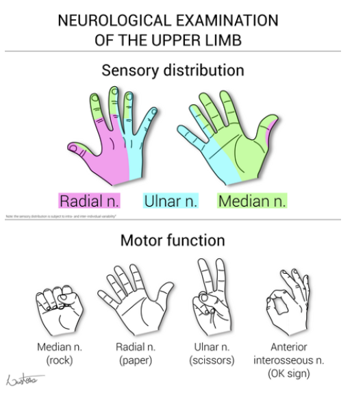
Ulnar nerve — most commonly injured; test finger abduction, sensation to small finger
Median nerve — test thumb opposition, sensation to index finger
Anterior interosseous nerve (AIN) — test flexion of thumb IP and index DIP ("OK sign")
Brachial artery — palpate radial and ulnar pulses; assess capillary refill
Skin integrity — open dislocations require urgent surgical management
Radiographic Evaluation
Alignment:
Anterior humeral line: drawn down the anterior surface of the humerus. It should intersect the middle 1/3 of the capitellum. If it does not, think distal humeral fracture.
Radiocapitellar line: drawn along the radial neck. It should always intersect the capitellum. If it does not, think radial head dislocation or subluxation. Check for an accompanying fracture, e.g. Monteggia fracture-dislocation
Effusion: visible posterior fat pad always indicates an elbow effusion
If there is an effusion, think acute intra-articular fracture. Elbow fractures may be occult on x-rays
Adult: radial head fracture
Child: supracondylar fracture
Bones: look specifically for common fractures
Radial head and neck fractures
Olecranon fracture
Cortex: trace the cortex of each bone
Distal humerus
Radial head, neck and shaft
Olecranon, coronoid process and ulnar shaft
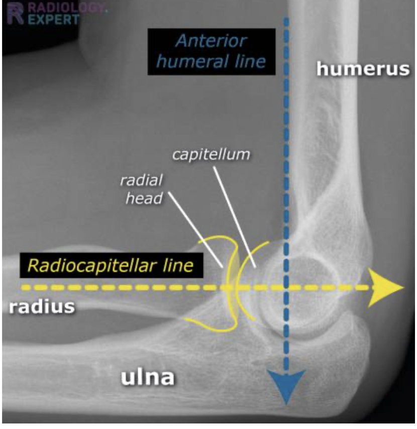
Normal Elbow Alignment
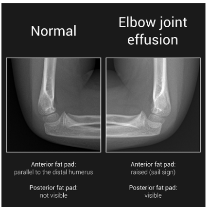
Joint Effusion
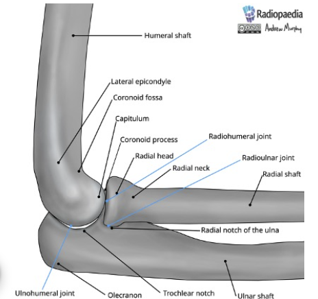
Bony Anatomy of Elbow
The Horii Circle of Soft Tissue Disruption
The model proposed that elbow dislocation results from posterolateral rotatory instability with soft tissue failure progressing sequentially from lateral to medial in a circle. The classic model describes sequential disruption from lateral to medial: Lateral collateral ligament (LUCL) Anterior capsule Posterior capsule Medial collateral ligament
Mechanism: Axial load + supination + valgus stress → the ulna rotates posterolaterally off the trochlea. However, studies have demonstrated that injury patterns are heterogeneous. Some dislocations show medially predominant injury ("reversed Horii circle")
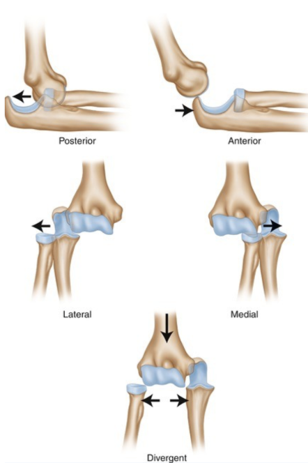
Takeaway: Always assess both medial and lateral stability after reduction, regardless of the assumed mechanism.
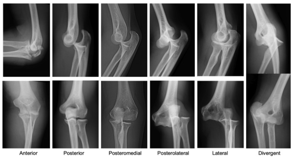
Classification – Direction & Complexity
By Direction:
Posterior / Posterolateral - Most common; olecranon displaced posteriorly
Anterior - Rare; direct blow to posterior olecranon
Medial / Lateral - Uncommon; pure translational displacement
Divergent - Very rare; radius and ulna separate from each other
By Complexity:
Simple: No associated fracture
Complex: Associated fracture(s)
Named Complex Patterns:
Terrible Triad — posterior dislocation + radial head fracture + coronoid fracture
Transolecranon fracture-dislocation — fracture through the olecranon with anterior displacement of the forearm
Monteggia variant — proximal ulna fracture with radial head dislocation
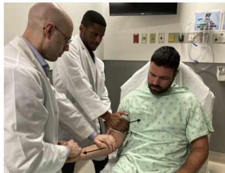
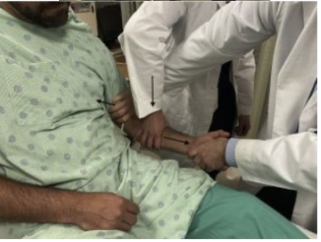
Traction-Countertraction
Reduction Techniques
General Principles
Perform under adequate procedural sedation and analgesia
Avoid forceful manipulation — gentle, controlled maneuvers reduce iatrogenic injury
An assistant is helpful but not always required
Posterolateral Dislocation:
Technique 1: Traction-Countertraction (Most Common)
Patient supine, elbow slightly flexed
Assistant stabilizes the humerus with both hands
Operator applies steady longitudinal traction to the forearm
While maintaining traction, gently flex the elbow
Apply anterior pressure over the olecranon to guide it distally and anteriorly
A palpable "clunk" confirms reduction
Technique 2: Leverage (No Assistant Needed)
Patient supine, arm on a flat surface
Operator grasps the proximal forearm with one hand and the distal humerus with the other
The forearm is used as a lever — the operator pushes down on the proximal forearm while lifting the distal humerus
This disengages the coronoid from the olecranon fossa without requiring forceful traction
Technique 3: Modified Stimson (Prone)
Patient prone with the affected arm hanging off the side of the bed
Gravity provides traction on the forearm
The operator applies gentle downward traction on the wrist and anterior pressure on the olecranon
Useful when sedation resources are limited
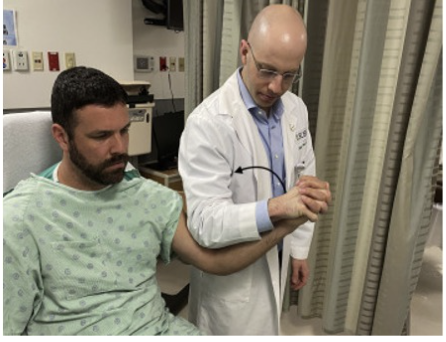
Levarage
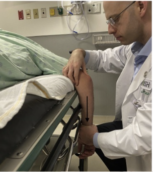
Modified Stimson (Prone)
Anterior Dislocation:
Technique: Modified Traction-Counteraction Technique
Posterior directed force applied on patient’s proximal forearm guides olecranon past distal humerus
Post-Reduction Assessment (Critical Step)
Move the elbow through a full arc of flexion-extension to determine the angle of re-dislocation (instability arc)
Apply varus and valgus stress at 30° of flexion to assess collateral ligament integrity
Assess forearm pronation/supination
Repeat neurovascular examination
Short term Immobilzation with posterior/long arm splint
Obtain post-reduction radiographs
Clinical Pearls
FOOSH is the most common mechanism of elbow dislocation
With any elbow dislocation, you need to check the shoulder and the wrist for injuries and fractures
Always perform and document a neurovascular exam before and after reduction—the ulnar nerve is the most commonly injured nerve.
Posterior fat pad sign indicates an occult intra-articular fracture until proven otherwise.
Terrible Triad = posterior elbow dislocation + radial head fracture + coronoid fracture is a highly unstable injury that usually requires operative management.
Reduce the elbow with gentle traction under adequate procedural sedation to minimize iatrogenic injury.
A reduced elbow is not necessarily a stable elbow, always repeat the neurovascular exam, obtain post-reduction radiographs, and assess elbow stability
Citations:
1. Gottlieb M. Managing Elbow Dislocations. Ann Emerg Med. 2022;80(5):460-464. doi:10.1016/j.annemergmed.2022.04.029. Available at: https://www.annemergmed.com/article/S0196-0644(22)00278-5/fulltext
2. O'Driscoll SW, Morrey BF, Korinek S, An KN. Elbow subluxation and dislocation: a spectrum of instability. Clin Orthop Relat Res. 1992;(280):186-197.
3. Morrey BF, An KN. Articular and ligamentous contributions to the stability of the elbow joint. Am J Sports Med.1983;11(5):315-319.
4. Morrey BF, An KN. Functional anatomy of the ligaments of the elbow. Clin Orthop Relat Res. 1985;(201):84-90.
5. O'Driscoll SW, Jupiter JB, King GJ, Hotchkiss RN, Morrey BF. The unstable elbow. J Bone Joint Surg Am.2000;82(5):724-738.
6. Reichert ILH, Ganeshamoorthy S, Aggarwal S, Arya A, Sinha J. Dislocations of the elbow—An instructional review. J Clin Orthop Trauma. 2021;21:101484. doi:10.1016/j.jcot.2021.101484. Available at: https://www.jsesreviewsreportstech.org/article/S2666-6391(21)00107-3/fulltext
7. Roberts JR, Custalow CB, Thomsen TW. Roberts and Hedges' Clinical Procedures in Emergency Medicine and Acute Care. 7th ed. Elsevier; 2019.
8. Tintinalli JE, Ma OJ, Yealy DM, et al., eds. Tintinalli's Emergency Medicine: A Comprehensive Study Guide. 9th ed. McGraw-Hill; 2020.
9. Orthobullets. Elbow Dislocation. Available at: https://www.orthobullets.com/trauma/1018/elbow-dislocation
10. Radiopaedia. Elbow Dislocation. Available at: https://radiopaedia.org/articles/elbow-dislocation?lang=us
11. Long B, Koyfman A. Elbow Dislocations. REBEL EM. Published November 18, 2021. Available at: https://rebelem.com/elbow-dislocations/