
Feet Pics for When the Foot Tricks: A Discussion of Base of the 5th Metatarsal Fractures
Written by: Hannah Weisman, MD PGY-1; Edited by: Timothy Khowong, MD, MSEd
Teaching Goals:
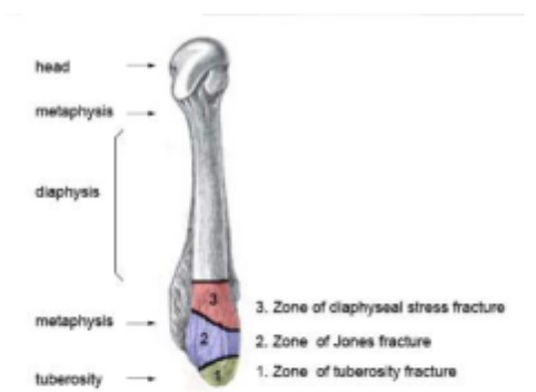
Define the 3 main types of base of the 5th metatarsal fractures
Learn how to recognize each fracture type
Understand the management of each type of fracture
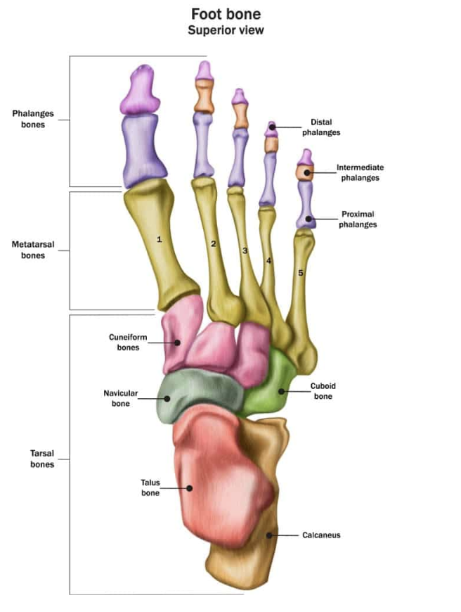
Bony Anatomy of the Foot:
Classifying Base of the 5th Metatarsal Fractures: The Lawrence and Botte 3-Zone Classification
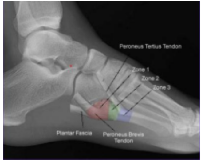
Physical Exam Features of Base of the 5th Metatarsal Fractures:
Pain, swelling, and ecchymosis over lateral border of forefoot
Point tenderness to palpation over the base of the 5th metatarsal
Pain with weight bearing or inability to bear weight
Zone 1: Pseudo-Jones Fracture:
Mechanism: Inversion injury. Caused when bony fragment is detached by ligament or other connective tissue
On imaging the fracture line extends into metatarsocuboid joint
Management:
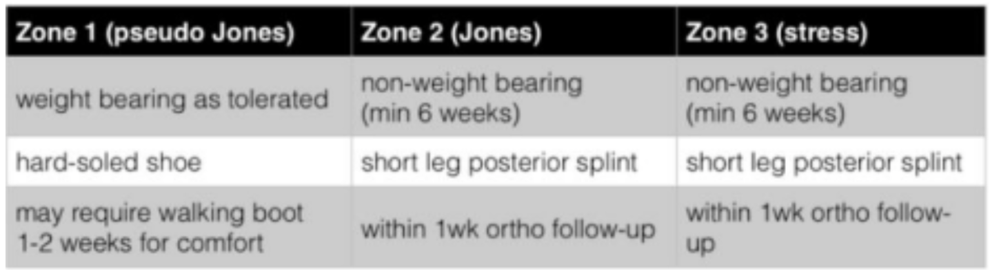
Weight bearing as tolerated
Discharge home in a hard-soled shoe
May require a walking boot for 1-2 weeks for comfort
Likely to heal on its own due to good perfusion
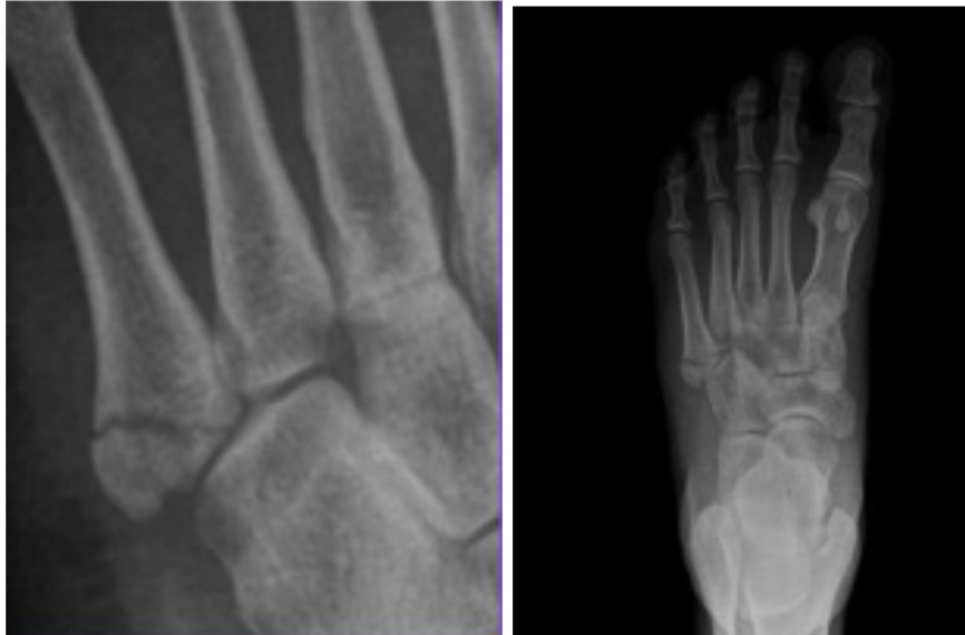
Pseudo-Jones Fractures
Zone 2: Jones Fracture
Mechanism: Forefoot adduction. Located at the Metaphyseal-diaphyseal junction (4th 5th metatarsal articulation)
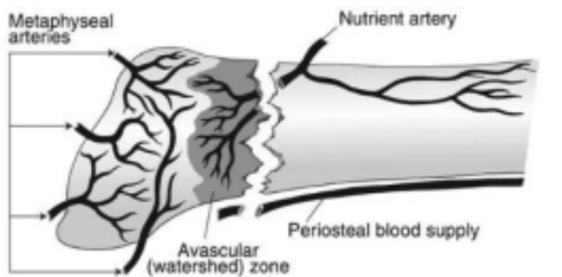
The proximal 5th metatarsal has unique blood supply with a watershed area at the metaphyseal-diaphyseal junction. Fractures in this area are at a high risk for non-union due to the fracture interrupting the already limited blood supply in this area
Management:
Non-weight bearing for 6 weeks
Short leg posterior splint, requires ortho follow up within 1 week
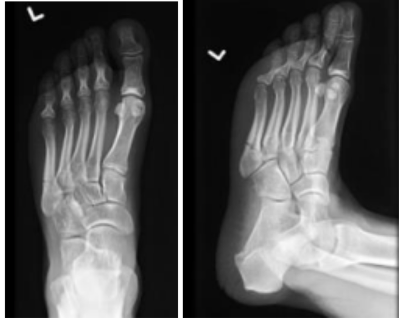
Left: Jones Fractures
Top: Watershed area at metaphyseal-diaphyseal junction.
Zone 3: Stress Fracture
Mechanism: Occurs from Repetitive microtrauma (stress fracture)
Proximal diaphyseal fracture (distal to 4th-5th metatarsal articulation), instead of well-defined fracture lines, stress injuries may have widened fracture lines with surrounding cortical thickening
Stress fractures typically occur within 1.5 cm of the metadiaphyseal junction, slightly distal to the site of a Jones fracture.
Associated with cavovarus foot deformities or sensory neuropathies
Management:
Non-weight bearing x6 weeks
Short leg posterior splint, requires ortho follow up within 1 week
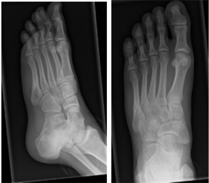
Stress fracture of 5th metatarsal
Take Home Points:
Location matters
These fractures are common with ankle sprains
Jones and stress fractures must be splinted with close ortho
follow-up!!
Summary:
Zone 1: Heal very well, non-union is rare, can resume activities as tolerated
Zone 2/3: Despite immobilization non-union can develop
High-caliber athletes are frequently treated with intra-medullary screw fixation
Non-union can lead to pain, difficulty ambulating, and possible need for surgery in the future
Given similar mechanism, have high index of suspicion for 5th metatarsal fractures in typical ankle sprains
References:
1.Fractures of the Base of the Fifth Metatarsal Bone: A Critical Analysis Review.
JBJS Reviews. 2021. Bušková K, Bartoníček J, Rammelt S.Review
2.Common Foot Fractures.
American Family Physician. 2024. Silver S, Williams E, Plunkett ML.Review
3.The Intraosseous and Extraosseous Vascular Supply of the Fifth Metatarsal: Implications for Fifth Metatarsal Osteotomy.
Foot & Ankle International. 2013. McKeon KE, Johnson JE, McCormick JJ, Klein SE.
4.Fractures of the Proximal Fifth Metatarsal: Selecting the Best Treatment Option.
The Journal of the American Academy of Orthopaedic Surgeons. 1995. Dameron TB.
5.Minimally displaced fracture involving the fifth metatarsal base.
Voio Radiology Database. VoioClinical Imaging
6.Fractures of the Proximal Fifth Metatarsal.
American Family Physician. 1999. Strayer SM, Reece SG, Petrizzi MJ.Review
7.National Athletic Trainers' Association Position Statement: Conservative Management and Prevention of Ankle Sprains in
Athletes.
Journal of Athletic Training. 2013. Kaminski TW, Hertel J, Amendola N, et al.Guideline
8.Bilateral Symptomatic Os Vesalianum Pedis: A Case Report.
The Journal of Foot and Ankle Surgery : Official Publication of the American College of Foot and Ankle Surgeons. 2011.
Dorrestijn O, Brouwer RW.Case
9.Lateral Foot Pain Due to Os Vesalianum Pedis in a Young Football Player; A Case Report and Review of the Current
Literature.
Skeletal Radiology. 2019. Aykanat F, Vincenten C, Cankus MC, Kose O, Sindel M.Review
10.Treatment Strategies for Acute Fractures and Nonunions of the Proximal Fifth Metatarsal.
The Journal of the American Academy of Orthopaedic Surgeons. 2000. Rosenberg GA, Sferra JJ.Review
11. Diagnosis and Management of Common Foot Fractures.
American Family Physician. 2016. Bica D, Sprouse RA, Armen J.Review
12. Return to Play and Fracture Union After the Surgical Management of Jones Fractures in Athletes: A Systematic Review
and Meta-Analysis.
The American Journal of Sports Medicine. 2021. Attia AK, Taha T, Kong G, et al.SR
13.Acute Fractures to the Proximal Fifth Metatarsal Bone: Development of Classification and Treatment Recommendations
Based on the Current Evidence.
Injury. 2012. Polzer H, Polzer S, Mutschler W, Prall WC.Review
14.Initial Assessment and Management of Select Musculoskeletal Injuries: A Team Physician Consensus Statement.
Medicine and Science in Sports and Exercise. 2024. Herring SA, Kibler WB, Putukian M, et al.Guideline
15. https://www.aafp.org/afp/2024/0200/common-foot-fractures (bony anatomy of foot)
16.Qureshi P, Dameron-Lawrence-Botte classification of proximal 5th metatarsal fractures. Reference article,
Radiopaedia.org (Accessed on 15 Jun 2026) https://doi.org/10.53347/rID-47848
17.https://coreem.net/core/jones-fractures/
18.Hanouneh S, Avulsion fracture of the 5th metatarsal tuberosity. Case study, Radiopaedia.org (Accessed on 15 Jun 2026)
https://doi.org/10.53347/rID-231106
19.https://radiologykey.com/lower-limb-iii-ankle-and-foot-2/
20.https://www.physio-pedia.com/Metatarsus_Adductus
21.Gaillard F, Dixon A, Stress fracture of the 5th metatarsal. Reference article, Radiopaedia.org (Accessed on 15 Jun 2026)
https://doi.org/10.53347/rID-7647
22.Mous A, Niknejad M, Jones fracture. Reference article, Radiopaedia.org (Accessed on 15 Jun 2026)
https://doi.org/10.53347/rID-1537
23.Yonso M, Jones fracture. Case study, Radiopaedia.org (Accessed on 15 Jun 2026) https://doi.org/10.53347/rID-99899