
What's in that scrotum?
Written by: Dr. Quinn Bushman
Edited by: Dr. Joann Hsu
Case:
A 47-year-old man presented to the ED with 2 months of worsening right-sided testicular swelling and 12 hours of worsening testicular pain. Pain acutely worsens before needing to urinate, and improves when the bladder is completely emptied. Denies any other urinary complaints, discharge, or rashes. His last bowel movement was this morning, and the patient continues to pass gas. Denies nausea, vomiting, or
fever.
Vitals: T 38.1ºC, BP 136/84, P 76 bpm, 97% on RA, RR 14
Physical Exam: Bilateral testicles are nontender, right scrotal sack is severely distended
but nontender; no erythema or tissue changes, no abdominal tenderness
Ultrasound:
Ultrasound was performed on the R scrotum
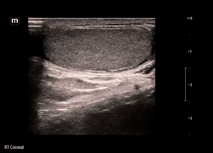
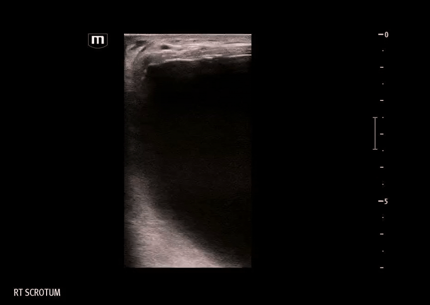
Findings on our bedside ultrasound:
The structure is anechoic - fluid-filled, appears to be simple fluid
The structure has no peristalsis, and there is a lack of typical bowel wall findings - probably not bowel or inguinal hernia
The structure does not seem to be within the tunica vaginalis - not a hydrocele, where the simple fluid collection would be seen surrounding the testicle itself
Comparison to other types of testicular pathologies:

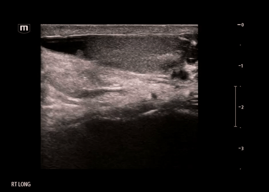
This image shows a hernia containing bowel - note the bowel wall layers, hyperechoic bowel contents

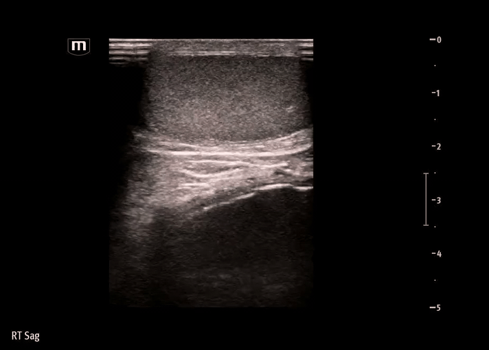
This image shows a hydrocele
In comparison, our image didn’t show typical bowel wall appearance but the swelling/mass was located within the scrotum, so what did the hernia contain?
A CT was obtained to further characterize the contents of the hernia:

Diagnosis: Inguinoscrotal bladder hernia
Inguinoscrotal bladder hernias are a rare form of sliding inguinal hernia in which a portion of the urinary bladder herniates through the inguinal canal and may extend into the scrotum.
They account for approximately 1–4% of all inguinal hernias, but large inguinoscrotal involvement (scrotal cystocele) is much less common and often underdiagnosed preoperatively.
Clinical presentation varies; many patients are asymptomatic, but classic symptoms include two-stage voiding (requiring manual compression of the scrotum to complete urination), lower urinary tract symptoms, or a scrotal mass that decreases in size after voiding.
Complications can include bladder incarceration, necrosis, hematuria, urinary tract infection, obstructive uropathy, and, rarely, acute renal failure.
Management:
Management is primarily surgical, with reduction or resection of the herniated bladder followed by tension-free herniorrhaphy.
The main goals are to preserve bladder function and avoid iatrogenic injury.
Large or complicated hernias may require urological consultation and tailored surgical planning.
Happy scanning!