The other EPSS
Written by: Dr. Seldon (Edwin) Davis
Edited by: Dr. Joann Hsu
The case:
35 YO M with pmh Crohn’s disease brought in by EMS for hematemasis, found to be pale and diaphoretic on scene, passed out, and became unresponsive. Patient was brought to the ED intubated.
Exam was limited since he was unresponsive and intubated but abdominal distension was noted.
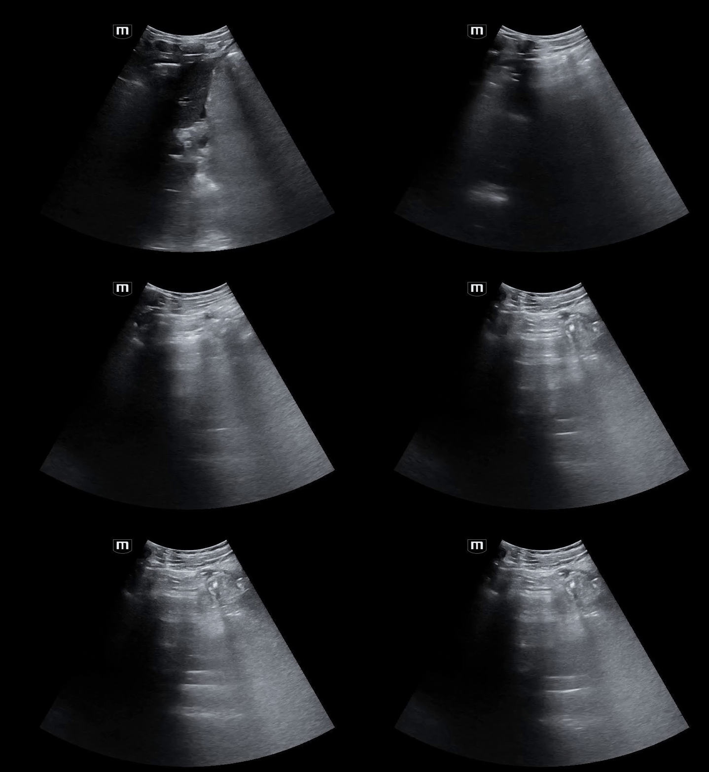
Bedside abdominal ultrasound in the RUQ showed:
RUQ with free fluid noted
Notice what looks like A lines coming in and out of the screen from the left, over the liver.
Again, the A lines over the liver.
Further imaging showed:
CT Abdomen/pelvis with IV contrast
Pneumoperitoneum with diffuse large and small bowel wall thickening,
most pronounced in the pelvic small bowel which is perforated
Moderate to large volume complex ascites, peritonitis, and multiple
loculated mesenteric gas and fluid-filled collections.
EPSS Basics
EPSS: Enhanced Peritoneal Stripe Sign
Enhanced (bright) echogenic line at the peritoneum
Concerning for pneumoperitoneum (free air within the abdomen)
EPSS is produced as the air interrupts echogenic return of the ultrasound waves.
The air causes a scattering of the ultrasound waves between the gas and soft tissue, producing a high-amplitude linear echo which leads to hyperechoic enhancement of the normal peritoneal stripe sign
So what is highlighted is the interface between gas and soft tissue!
EPSS and other Ultrasonographic Findings concerning for Pneumoperitoneum
EPSS
Reverberation Artifact (A-lines in the Abdomen)
Comet-Tail Reverberatory Artifacts
Air Bubbles in Ascitic Fluid
EPSS is a classic finding concerning for pneumoperitoneum, however in the setting of substantial pneumoperitoneum, A-Lines can be visualized within the abdomen
***** Not to be confused with EPSS/E-Point Septal Separation, measurement of the of anterior Mitral Valve leaflet movement used as an estimate for Left Ventricular Ejection Fraction
Tips and Tricks
Use a Linear/Curvilinear Transducer
Look for signs of air being intra-luminal within the bowel (peristalsis, dirty shadowing)
Look in the RUQ
Anterior aspect of liver is adjacent to the anterior abdominal wall
Not occupied by bowel, and should be free from bowel gas
Scissor Maneuver
Slight Pressure to ant. Abd --> free intraperitoneal air expelled from region anterior to liver to other parts of peritoneal cavity --> reverberation artifact becomes less prominent
Then when pressure on distal end of probe is released, maintain contact with probe and skin surface --> free gas returns to the area and the A-lines become more prominent
In real time the repeated maneuver appears like opening and closing of scissors
Literature
Perforated Viscus associated w/ pneumoperitoneum, is a life-threatening etiology of acute abdominal pain in the ER
Literature supports POCUS as a useful adjunct and tool to rapidly diagnose and expedite management
Back to the case:
The patient was taken to the OR and after an extensive hospital course, he was discharged to rehab for further recovery.
Happy scanning!
References
Bacci M, Kushwaha R, Cabrera G, et al. (June 08, 2020) Point-of-Care Ultrasound Diagnosis of Pneumoperitoneum in the Emergency Department. Cureus 12(6): e8503. doi:10.7759/cureus.8503
Carroll D, Elfeky M, Shah V, Peritoneal stripe sign (pneumoperitoneum). Reference article, Radiopaedia.org (Accessed on 29 Sep 2024) https://doi.org/10.53347/rID-65535
Chao, A., Gharahbaghian, L., & Phillips, P. (2014, December 23). Diagnosis of Pneumoperitoneum with Bedsite Ultrasound. Western Journal of Emergency Medicine, 16(2).
Indiran, V., Vinoth Kumar, R. & Jefferson, B. Enhanced peritoneal stripe sign. Abdom Radiol 43, 3518–3519 (2018). https://doi.org/10.1007/s00261-018-1628-7
Kricun, B. J., & Horrow, M. M. (2012, June). Pneumoperitoneum. Ultrasound Quarterly, 28(2)
Muradali D, Wilson S, Burns PN, Shapiro H, Hope-Simpson D. A specific sign of pneumoperitoneum on sonography: enhancement of the peritoneal stripe. AJR Am J Roentgenol. 1999 Nov;173(5):1257-62. doi: 10.2214/ajr.173.5.10541100. PMID: 10541100.


