
Ovarian Torsion
Written by: William Chan, MD. Edited by: Jeff Greco, MD
Overview
Ovarian torsion is one of the many can’t miss diagnoses in the emergency room. It refers to a complete or partial rotation of the ovary within the adnexa. It involves the twisting of the ovary on its ligamentous support. To review some quick anatomy, the ovary is supported by the infundibulopelvic (suspensory) ligament which attaches to the pelvic side wall and the utero-ovarian ligament which attaches the ovary to the uterus. The blood supply of the ovary is held within these ligaments, so the twisting of these ligaments can lead to obstructed flow. Although most occur in females of childbearing age, approximately 15% of cases occur in pediatric patients and approximately 15% of cases occur post-menopausal. Don’t forget that ovarian torsion can still occur during pregnancy. Putting your ultrasound (US) skills to use is a great way to help you make this diagnosis. US doppler to look for arterial flow is one of the most popular US signs to look for in torsion cases. However, the purpose of this post is to take a closer look at some of the other signs of ovarian torsion.
Enlarged Ovary
An ovary with dimensions greater than 2x3x4 cm is considered enlarged and can be a sign of torsion. Below is an example of an enlarged ovary, with the dimensions of approximately 4x5x7 cm.
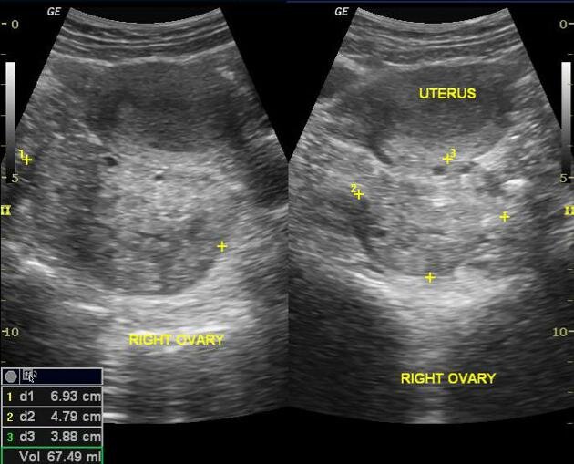
Midline ovary
As an ovary torses, the length of the ligaments supporting it can shorten. This can cause the position of the ovary in the abdominal cavity to shift more medially. The image below is a transabdominal ultrasound that demonstrates this change in position. On the image right, is the left ovary that is grossly normal. On the image left, is the right ovary that is enlarged and has shifted medially and is now adjacent to the left ovary.


Ovarian mass
More than 80% of patients with ovarian torsion had an ovarian mass of 5 cm or greater. Torsion can happen with masses of any size and has been reported in cases with masses of 1 to 30 cm. The image below shows an approximately 3 cm hemorrhagic cyst within an ovary.
Pelvic free fluid
Finding pelvic free fluid on US can be a sign of ovarian torsion. In this clinical setting, it can possibly represent hemorrhage from the ovary or inflammation from the ischemia of the ovary. In the image below, the anechoic area to the left of the ovary is free fluid.

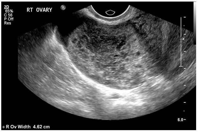
Peripheralization of follicles
As the ovary becomes more edematous, the follicles are pushed to the periphery of the ovary. In the image below, the ovary is the ovaloid structure that is surrounded by the hyperechoic capsule. The follicles are the hypoechoic structures adjacent to the capsule on the right side of the image.

Whirlpool sign
Whirlpool sign is the swirling appearance of the vessels in the twisted pedicle. In grey-scale US, it is typically an echogenic round mass with multiple hypoechoic, target-like stripes. With doppler sonography, the coiled vessels can be better appreciated. In the left image below, there is a circular mass within the arrows with hypoechoic and hyperechoic textures. In the right image below, the visualization of the coiled vessels within the mass can be appreciated with the assistance of doppler.
References
Chang HC, Bhatt S, Dogra VS. Pearls and pitfalls in diagnosis of ovarian torsion. Radiographics. 2008;28(5):1355-1368. doi:10.1148/rg.28507513
Dixon A. Ovarian torsion: Radiology Reference Article. Radiopaedia Blog RSS. https://radiopaedia.org/articles/ovarian-torsion?lang=us. Accessed January 14, 2021
Guile SL, Mathai JK. Ovarian Torsion. In: StatPearls. Treasure Island (FL): StatPearls Publishing; November 20, 2020.
Huang C, Hong MK, Ding DC. A review of ovary torsion. Ci Ji Yi Xue Za Zhi. 2017;29(3):143-147. doi:10.4103/tcmj.tcmj_55_17
Ormsby EL, Geng J, McGahan JP, Richards JR. Pelvic free fluid: clinical importance for reproductive age women with blunt abdominal trauma. Ultrasound Obstet Gynecol. 2005;26(3):271-278. doi:10.1002/uog.1981
Sintim-Damoa A, Majmudar AS, Cohen HL, Parvey LS. Pediatric Ovarian Torsion: Spectrum of Imaging Findings. Radiographics. 2017;37(6):1892-1908. doi:10.1148/rg.2017170026
Themes UFO. 26 25-year-old female presented with nausea, vomiting, and left abdominal pain. Radiology Key. https://radiologykey.com/26-25-year-old-female-presented-with-nausea-vomiting-and-left-abdominal-pain/. Published February 19, 2017. Accessed January 14, 2021