
POCUS for Diverticulitis
Written by: Justin Wang, MD. Edited by: Jeff Greco, MD.
Overview
Abdominal pain is one of if not the most common chief complaint presenting to the emergency room. Most people with significant enough findings will end up requiring a CT scan for complete evaluation of possible complications. However, CT scans aren’t completely benign as it does expose patients to radiation and can significantly prolong disposition while you wait for the scan. In patients we suspect with diverticulitis, we may be able to use the point of care ultrasound to save patients from unnecessary radiation and as an easy and reliable way to get them appropriate treatment faster.
Evidence

Primary Outcome - Test characteristics of POCUS compared to CT for diagnosis of diverticulitis
Sensitivity 0.92 (0.88–0.96)
Specificity 0.97 (0.94–0.99)
LR+ 30.67
LR- 0.08
Average US time: ~5 minutes
Primary Outcome - Test characteristics of POCUS compared to CT for diagnosis of diverticulitis
Sensitivity 92.7%
Specificity 90.9%
Thickened bowel wall appears to be most sensitive criteria
Technique
Curvilinear or linear probe

Start in the location of greatest pain. Progress with lawnmower technique if no diverticulitis identified

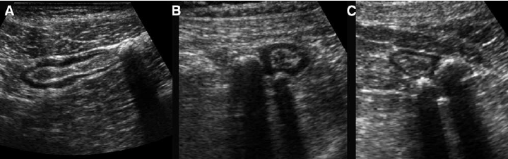
General consensus criteria for positive diagnosis
Diverticula: hypoechoic domed structure extending off of the colon
Bowel wall edema >4- 5 mm surrounding a diverticula
Enhancement/inflammation of pericolonic fat
Sonographic tenderness to palpation
Source: https://www.criticalcare-sonography.com/2019/03/03/diverticulitis/


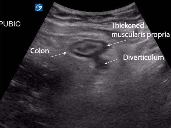
Sigmoid colon and hyperechoic pericolic fat surrounding diverticulum
Source: https://www.ultrasoundcases.info/diverticulosis---diverticulitis-3605/

Inflamed perienteric fat surrounding a diverticulum
Source: https://www.ultrasoundcases.info/diverticulosis---diverticulitis-531/

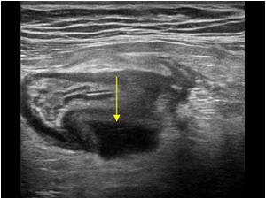
Measurement of bowel wall edema
Source: Gasche C, et al.
Other possible findings
Fecalith: central shadowing echogenicity

“Sonographic features of uncomplicated diverticulitis: diverticula appear as bright “ear” out of the bowel wall (a); a central shadowing echogenicity may indicate the presence of fecalith (b).”
Source: https://theultrasoundjournal.springeropen.com/articles/10.1186/2036-7902-5-S1-S5
“( A , B , C ) Sigmoid diverticulosis in three asymptomatic patients. The fecalith-filled diverticula are recognized as strongly reflective, round structures casting an acoustic shadow and localized at the outer contour of the empty sigmoid. The thin wall of the diverticulum, consisting of mucosa only, is not separately visible.”
Source: Puylaert, Julien. (2003). Ultrasonography of the acute abdomen: gastrointestinal conditions. Radiologic clinics of North America. 41. 1227-42, vii.
Complicated diverticulitis: Intramural/pericolic abscess seen as an anechoic collection of air/debris

“Sonographic features of complicated diverticulitis: the images show the presence of diveticula, thickening of the bowel wall and pericolic fluid (a,b)”
Source: https://theultrasoundjournal.springeropen.com/articles/10.1186/2036-7902-5-S1-S5
Mural abscess
Source: https://www.ultrasoundcases.info/diverticulosis---diverticulitis-6835/

Paracolic abscess caused by diverticulitis, effectively walled-off by large masses of inflamed fat, representing mesentery and omentum. The abscess eventually evacuated completely, and the patient recovered without surgery.
Source: Puylaert, Julien. (2003). Ultrasonography of the acute abdomen: gastrointestinal conditions. Radiologic clinics of North America. 41. 1227-42, vii.
Summary
Acute uncomplicated diverticulitis can be accurately and quickly diagnosed in the appropriate patient with point of care ultrasound
Consider sparing patients unnecessary radiation and wait times and pick up your trusty probe instead!
References
Mazzei, M.A., Cioffi Squitieri, N., Guerrini, S. et al. Sigmoid diverticulitis: US findings. Crit Ultrasound J 5, S5 (2013). https://doi.org/10.1186/2036-7902-5-S1-S5
Carbonatto, Genevieve. Diverticulitis. Critical Care Sonography. Published on March 3, 2019. Accessed on November 20, 2020. Available at https://www.criticalcare-sonography.com/2019/03/03/diverticulitis/
Michael Prats. The Accuracy of POCUS for Diverticulitis. Ultrasound G.E.L. Podcast Blog. Published on November 09, 2020. Accessed on November 20, 2020. Available at https://www.ultrasoundgel.org/101.
Cohen A, Li T, Stankard B, Nelson M. A Prospective Evaluation of Point-of-Care Ultrasonographic Diagnosis of Diverticulitis in the Emergency Department. Ann Emerg Med. 2020 Jul 8:S0196-0644(20)30365-6. doi: 10.1016/j.annemergmed.2020.05.017. Epub ahead of print. PMID: 32653332.
Nazerian P, Gigli C, Donnarumma E, de Curtis E, Bribani A, Lanzi S, Rovida S, Magazzini S, Grifoni S, Perani C. Diagnostic Accuracy of Point-of-Care Ultrasound Integrated into Clinical Examination for Acute Diverticulitis: A Prospective Multicenter Study. Ultraschall Med. 2020 Jul 20. English. doi: 10.1055/a-1161-0780. Epub ahead of print. PMID: 32688404.
Puylaert, Julien. (2003). Ultrasonography of the acute abdomen: gastrointestinal conditions. Radiologic clinics of North America. 41. 1227-42, vii.
Gasche C, Moser G, Turetschek K, et alTransabdominal bowel sonography for the detection of intestinal complications in Crohn’s diseaseGut 1999;44:112-117.