Navigating Lunate and Perilunate Dislocations in the Emergency Department
Written by: Andres Somoza, MD; Edited by: Timothy Khowong, MD, MSEd
Emergency Evaluation, Imaging Patterns, and Pitfalls
Lunate and perilunate dislocations represent high-risk, high-energy carpal injuries that carry severe long-term functional consequences if recognition is delayed. Despite their critical nature, these injuries are frequently missed on initial evaluation in the emergency department (ED). Understanding carpal alignment, recognizing subtle radiographic signs, and executing proper acute management are essential skills for any clinician managing wrist trauma.
Functional Anatomy of the Wrist
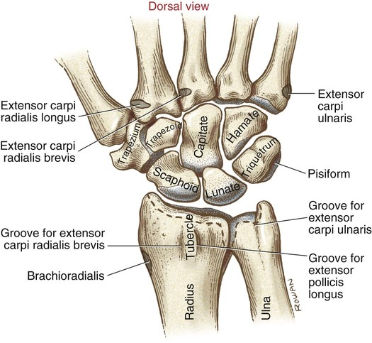
To identify subtle disruptions, clinicians must systematically understand normal carpal anatomy. The wrist comprises eight carpal bones arranged in two distinct rows, stabilized by a dense network of intrinsic and extrinsic ligaments:
Proximal Row: Scaphoid, lunate, triquetrum, and pisiform.
Distal Row: Trapezium, trapezoid, capitate, and hamate.
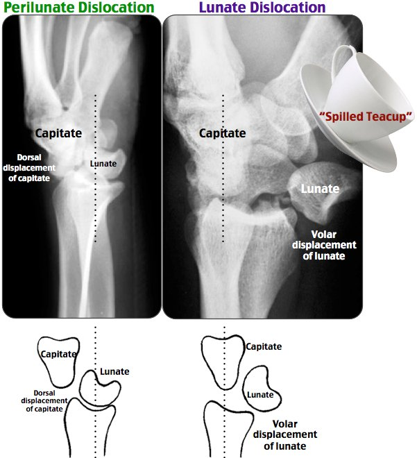
The lunate acts as the anatomical keystone of the wrist, articulating proximally with the radius and distally with the capitate. Maintaining the collinear alignment of the radius, lunate, capitate, and third metacarpal is central to preserving normal carpal mechanics.
Demographics and Mechanism of Injury
Lunate and perilunate dislocations typically stem from high-energy trauma. Common mechanisms include:
Motor vehicle accidents (MVAs) and motorcycle collisions
Pedestrian versus auto accidents and bicycle collisions
Falls from significant height (FOOSH with forced extension and ulnar deviation)
High-impact sports trauma and physical assaults
These injuries occur predominantly in young male patients. Paresthesias in the median nerve distribution (numbness over digits 1–3 and weakness in thumb opposition) are present in approximately 48% of cases at initial presentation.
The Diagnostic Challenge: Why Are These Injuries Missed?
Up to 25–30% of lunate and perilunate dislocations are missed during the initial emergency department visit. Clinicians often face diagnostic traps:
Subtle Gross Deformity: Significant soft tissue swelling can easily hide marked carpal bone displacement.
Distracting Injuries: High-energy trauma frequently involves poly-trauma, drawing clinical focus away from the wrist.
Radiographic Inexperience: Emergency practitioners may fail to perform a systematic assessment of carpal alignment on lateral views.
Misdiagnosis: Pain and swelling are easily misattributed to a severe wrist sprain or Triangular Fibrocartilage Complex (TFCC) injury.
Radiographic Evaluation and Alignment Rules
Source: Wikipedia
Standard initial imaging includes PA, lateral, and oblique radiograph views. In complex trauma settings, CT scanning may be utilized for precise operative planning.
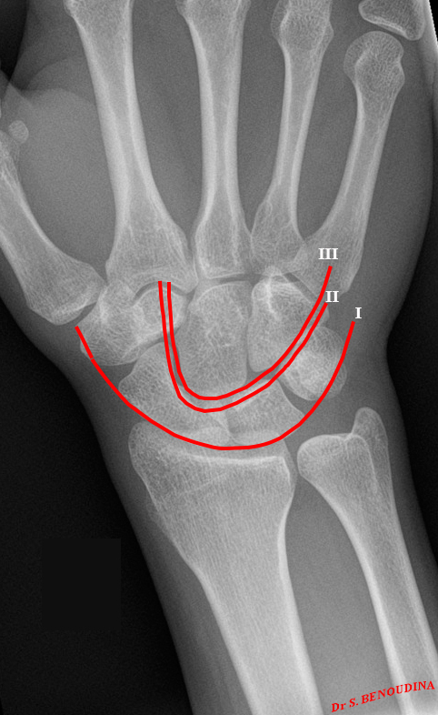
On the PA radiograph, carpal alignment is evaluated using Gilula's three carpal arcs:
Arc I: Smooth curve along the proximal convexities of the proximal carpal row (scaphoid, lunate, triquetrum).
Arc II: Smooth curve along the distal concavities of the proximal carpal row.
Arc III: Smooth curve along the proximal margins of the distal carpal row (capitate and hamate).
Any step-off, disruption, or overlap among these arcs signals significant ligamentous disruption or fracture-dislocation. The trans-scaphoid perilunate fracture-dislocation represents the most common pattern, where the injury force propagates through the scaphoid body rather than the scapholunate ligament.
Lateral View Assessment: Collinear Alignment
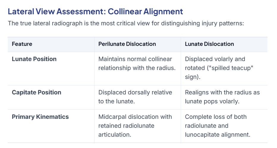
The true lateral radiograph is the most critical view for distinguishing injury patterns:
Source: Rosh Review
Emergency Management and Reduction Considerations
Prompt consultation with an orthopedic surgeon is mandatory upon recognizing a carpal dislocation pattern. Emergency reduction should be performed promptly to relieve tension on surrounding neurovascular structures.
Procedural Sedation vs. Regional Nerve Blocks
While hematoma blocks, wrist blocks, or Bier blocks offer effective localized analgesia, procedural sedation is strongly preferred for lunate and perilunate dislocations due to two critical factors:
Adequate Muscle Relaxation: Significant manual traction is required to reduce a dorsally displaced capitate or volarly rotated lunate; regional blocks frequently fail to provide sufficient muscle relaxation.
Preserving Median Nerve Assessment: Median nerve compression is present in 48% of initial presentations and eventually requires carpal tunnel release in 52% of cases. Nerve blocks temporarily mask evolving neurologic deficits, whereas procedural sedation allows immediate post-reduction neurological reassessment.
Reduction Techniques
Dorsal (Perilunate) Dislocations: Reduced using longitudinal traction combined with direct dorsal pressure over the capitate while guiding the wrist into flexion.
Volar (Lunate) Dislocations: Reduced using longitudinal traction paired with palmar pressure over the rotated lunate combined with capitate manipulation.
Surgical Management and Long-Term Complications
Definitive treatment almost universally requires operative intervention:
Open Reduction and Internal Fixation (ORIF): Represents the primary surgical strategy in approximately 56% of cases.
Salvage Procedures: In chronic missed dislocations, proximal row carpectomy (PRC) may be performed, provided the articular surfaces of the lunate fossa and proximal capitate are preserved. Total wrist arthrodesis or arthroplasty remains reserved for severe, complex cases.
Expected Complications
Even with timely reduction and surgical fixation, outcomes can be guarded:
Posttraumatic Arthritis: Develops in up to 90% of patients, with radiographic degeneration noted in 79% of long-term follow-ups.
Median Nerve Injury: Risk of permanent sensory or motor deficits from prolonged carpal tunnel compression.
Avascular Necrosis (AVN): Interrupted nutrient blood supply to the lunate or scaphoid fragments.
Functional Deficits: Chronic stiffness, decreased range of motion (ROM), reduced grip strength, and persistent wrist pain.
Disposition Criteria
Discharge directly from the ED is never appropriate for acute lunate or perilunate dislocations. All patients require hospital admission and urgent orthopedic surgery follow-up due to:
High risk of acute post-reduction re-displacement
Impending need for urgent operative fixation
High threat of progressive median nerve compromise
Complex pain management and monitoring requirements
References
Kannikeswaran N, Sethuraman U. Lunate and Perilunate Dislocations. Pediatric Emergency Care. 2010;26(12):921-4.
Scalcione LR, Gimber LH, Ho AM, et al. Spectrum of Carpal Dislocations and Fracture-Dislocations: Imaging and Management. AJR. American Journal of Roentgenology. 2014;203(3):541-50.
Patel AU, Thiara J, Rivas Z, Hansen SL. Descriptive Epidemiology and Management of Perilunate Injuries. Annals of Plastic Surgery. 2024;93(4):447-450.
Perron AD, Brady WJ, Keats TE, Hersh RE. Orthopedic Pitfalls in the ED: Lunate and Perilunate Injuries. The American Journal of Emergency Medicine. 2001;19(2):157-62.
Goodman AD, Harris AP, Gil JA, et al. Evaluation, Management, and Outcomes of Lunate and Perilunate Dislocations. Orthopedics. 2019;42(1):e1-e6.
Stanbury SJ, Elfar JC. Perilunate Dislocation and Perilunate Fracture-Dislocation. The Journal of the American Academy of Orthopaedic Surgeons. 2011;19(9):554-62.
Kozin SH. Perilunate Injuries: Diagnosis and Treatment. The Journal of the American Academy of Orthopaedic Surgeons. 1998;6(2):114-20.
Heineman N, Do DH, Golden A. Carpal Dislocations. The Journal of Hand Surgery, European Volume. 2023;48(2_suppl):11S-17S.
Kapoor G, Heire P, Turmezei T, Chojnowski A, Toms AP. Perilunate Injuries: Biomechanics, Imaging, and Classification. Clinical Radiology. 2020;75(2):81-87.
Obert L, Loisel F, Jardin E, Gasse N, Lepage D. High-Energy Injuries of the Wrist. Orthopaedics & Traumatology, Surgery & Research. 2016;102(1 Suppl):S81-93.
Budoff JE. Treatment of Acute Lunate and Perilunate Dislocations. The Journal of Hand Surgery. 2008;33(8):1424-32.
Sawardeker PJ, Kindt KE, Baratz ME. Fracture-Dislocations of the Carpus: Perilunate Injury. The Orthopedic Clinics of North America. 2013;44(1):93-106.