
ANKLE FRACTURES: A Joint Effort Gone Wrong
Written by: Marlene Louro, DO; Edited by: Timothy Khowong, MD, MSEd
Let’s meet our patient:
A 51 year-old male presents to the ED for left foot pain after a mechanical fall. Patient states he slipped on a thin paper-like item and slipped having his left foot slide forward, falling onto his bottom. He was originally able to ambulate but now is having difficulty secondary to pain.
Vitals: Within normal limits
Physical Exam: Left ankle is edematous and ecchymotic at lateral and medial malleoli with TTP. No TTP over the tibia/fibula or foot, +2 DP pulses, limited ROM of left ankle
We’ll come back to this case at the end but in the meantime, what imaging would you want to order and what are some differentials?
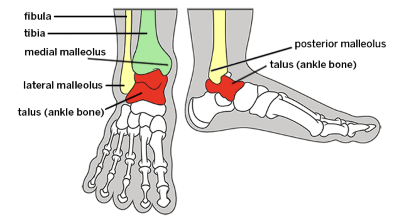
What makes up the ankle?
There are three bones that make up the ankle: tibia, fibula and talus. The lateral and posterior malleoli are part of the fibula bone. The medial malleolus is part of the distal tibia.The mortise of the ankle is the socket where the distal tibia and fibula meet the talus.
The ankle is also made up of many ligaments. On the lateral aspect of the ankle we have the anterior talofibular ligament, calcaneofibular ligament and the posterior talofibular ligament. On the medial side of the ankle, we have the deltoid ligament. It is made up of four ligaments and is what provides most stability for the ankle.
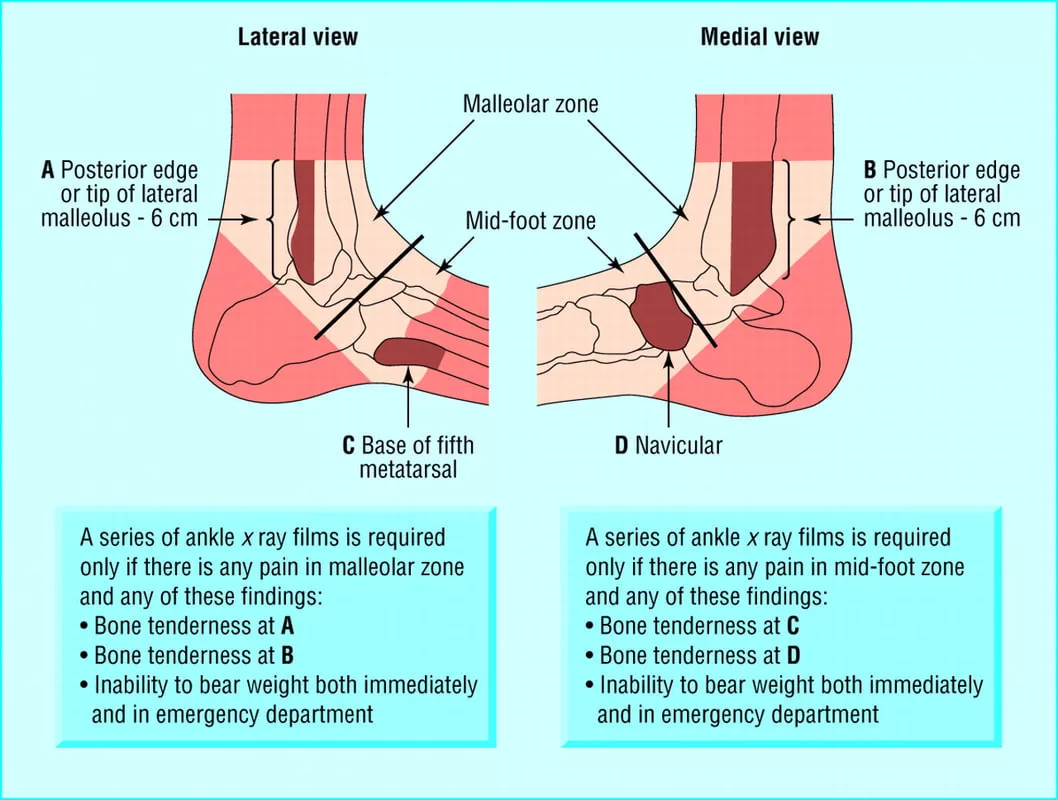
How can the ankle become injured?
Rotational injury: ankle is twisted, turned or rolled. Usually caused by walking, running or playing sports.
High Force Impact: usually due to a fall or motor vehicle accident.
Stress Fracture: occurs most often in the tibia and fibula. Usually due to a person beginning a new activity that involves significant impact on the foot like hiking or running. Or, a person who quickly increases their activity such as occasionally jogging to suddenly training for a marathon.
Use the Ottawa Ankle Rules to help decide when X rays are needed!
Let’s review how to interpret X Rays of the ankle:
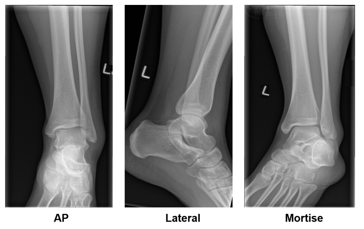
You will need to obtain 3 views of the ankle and in some cases, will need to obtain a fourth. The three main views include
AP
Lateral
Mortise
The mortise view is a modified AP view with 10-20 degrees of internal rotation of the foot so that both the medial and lateral malleoli are in horizontal plane and the joint visualization is optimized.
When reading these x rays, you want to take note of a few important things:
Look at all the contours of the bones to ensure there is no fracture
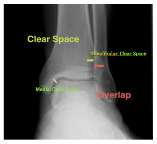
In a normal ankle x ray, the tibiofibular overlap should be >6mm.
In a normal ankle x ray, the medial clear space should be ≤4mm.
In a normal ankle x ray, the tibiofibular clear space should be <6mm.
What is the stress view?
In some cases, a stress view of the ankle is required to determine if the ankle is stable or unstable. Specifically, it is looking to see if there is a tear in the deltoid ligament, rendering the ankle unstable.
How to obtain a manual stress view:
Internally rotate the leg, dorsifex and externally rotate the foot (big toe to the sky)
The mortise of the ankle should uniformly be <4mm. If there is widening of medial clear space more than 4mm, there is injury to the deltoid ligament and the ankle is unstable. Ortho must be consulted!
Now that we’ve got the basics down, let’s talk about the different types of ankle fractures!
Lateral Malleolus Fracture
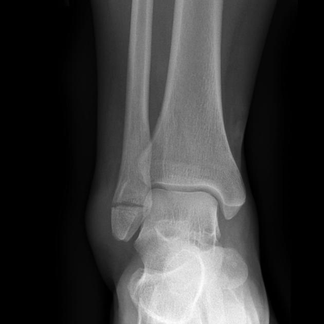
Lateral Malleolus fracture is described using the Weber Classification:
Weber A: Fracture of the fibula is below the tibiofibular syndesmosis.
These are stable fractures
Patients can be placed in a short leg posterior splint or boot. They are weight bearing.
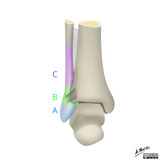
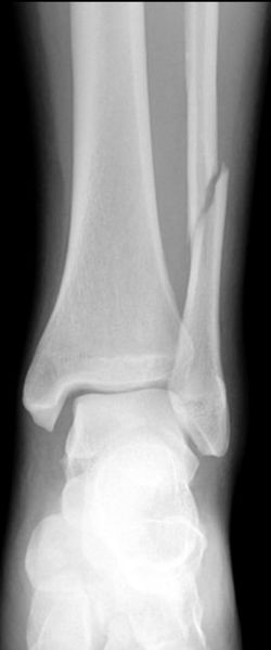
Weber C: Fracture above the tibiofibular syndesmosis.
Always unstable
Can still obtain stress views to determine degree of instability.
Consult ortho for reduction, splinting and OR.
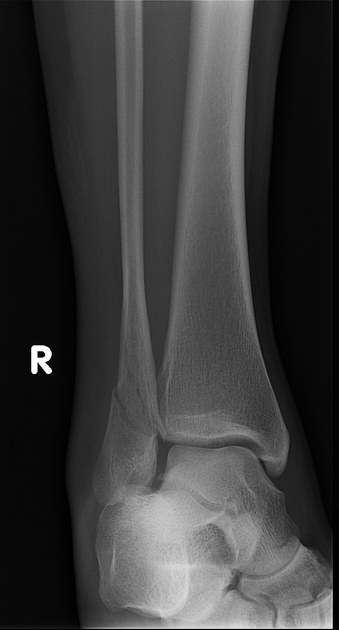
Weber B: Fracture at the level of the tibiofibular syndesmosis.
Can be stable or unstable → must do stress view x rays to determine
If stable, short leg posterior splint or boot.
If unstable, consult ortho for reduction, splinting and OR.
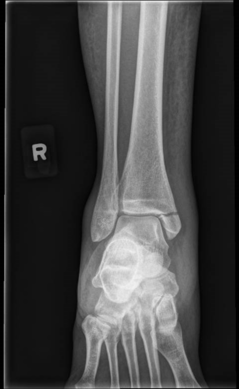
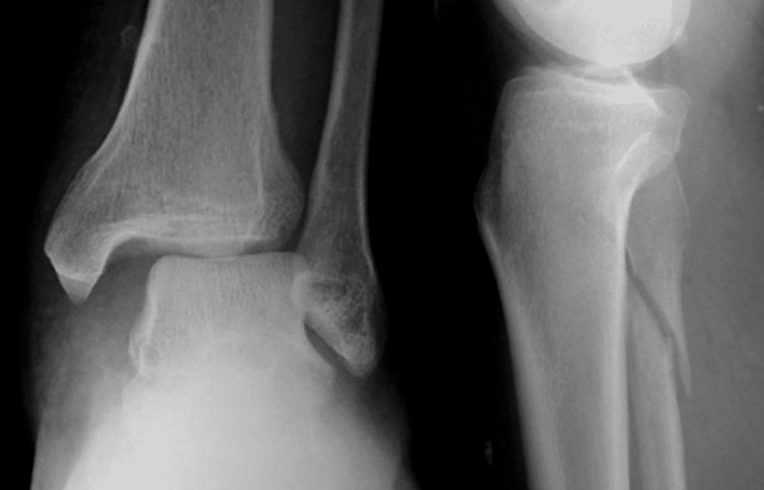
Maisonneuve Fracture
Spiral fracture of the proximal fibula w/ unstable ankle injury and disruption of distal tibiofibular syndesmosis.
Can be associated with a medial malleolus fracture or injury to the ligaments on the medial aspect of the ankle as well as posterior malleolus fracture.
If there is unexplained increase in medial clear space or tibiofibular clear space is widened, consider maisonneuve fracture.
Usually due to external rotation with pronation of the foot.
Consult ortho, patient needs surgery.
Isolated Medial Malleolar Fracture
Usually stable
NWB for 4-6 weeks
If talar shift is present
ORIF
Make sure to palpate the tib-fib and obtain tib-fib x rays because…. Maisonneuve Fractures!
Trimalleolar Fracture
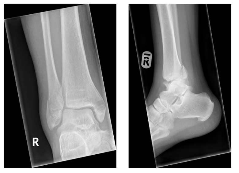
Fracture of medial, lateral and posterior malleoli.
Usually caused by rotational injury, MVCs, or falls.
Always unstable.
Consult ortho, needs ORIF.
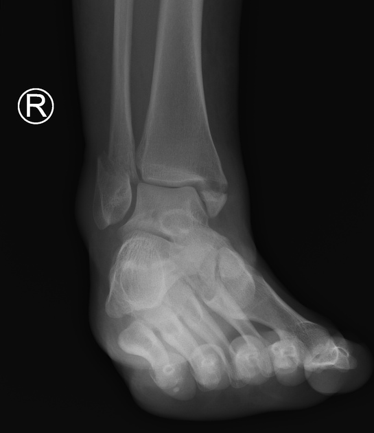
Bimalleolar Fracture
Fracture of the lateral and medial malleoli
Almost always unstable
Consult ortho, patient needs surgery
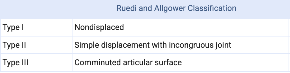
Pilon Fracture aka Tibital Plafond Fracture:
Fracture of the distal end of the tibia
Usually due to high energy axial load
Associated with comminution and intraarticular fractures
75% have associated fibula fractures
Will require CT scan
Classified using Ruedi and Allgower Classification
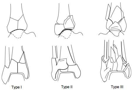
Most cases require temporary external fixation followed by delayed open reduction internal fixation
If non-operative (no articular surface displacement), patient will require long leg cast for 6 weeks w/ close orthopedic follow up
Back to our case:
Our patient had a trimalleolar fracture! Ortho was consulted- they reduced the fracture, placed the patient posterior AO splint and was scheduled for ORIF