Old McConnell Had a Fall
Written by: Dr. Haroon Karabay
Edited by: Dr. Joann Hsu
The case:
93 y/o f with PMH of HTN, HLD BIBEMS from home for shortness of breath.
Patient is A&Ox1 ( to self ) and reports that she had a fall an unknown time ago and is complaining of left lower leg pain.
Unable to obtain further history at the time.
Vitals: BP 121/78, T 36.8 C, SpO2 on RA 94%, RR 22
Physical Exam:
Saturating at 94% on 6L nasal cannula, mildly tachypneic
Clear lung sounds
Normal cardiac exam including RRR, no MRR.
No signs of external trauma
Bilateral distal lower extremity swelling and tenderness to palpation.
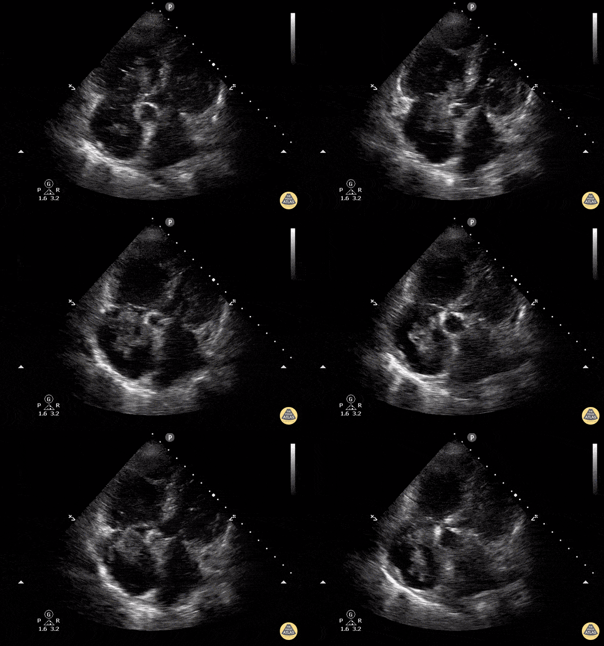
Given the vitals and physical exam you have increased concern for acute PE. You perform bedside cardiac point-of-care ultrasound (POCUS) and note the below finding in your apical 4-chamber view:
What do you see?
Background:
Right heart strain (more precisely, right ventricular strain) is a term given to describe the presence of right ventricular dysfunction in the setting of acute or chronic pathology.
Right heart strain often occurs because of pulmonary arterial hypertension (a chronic process).
One very common cause of acute right ventricular strain is pulmonary embolism.
The reported sensitivity and specificity of echocardiography in demonstrating right heart dysfunction are 56% and 42%, respectively.
In the emergency department, it may be difficult to definitively measure right heart strain but we can use bedside ultrasound to look for evidence of RV strain, particularly acute RV strain:
McConnell’s sign
Increased RV:LV ratio
Direct visualization of an RV thrombus
Abnormal septal motion (D sign seen in the parasternal short axis view)
Decreased TAPSE
Pulmonary arter mid-systolic notching
60/60 sign
We will not go into all of these today, but there was one particular finding in this case: McConnell’s sign!
McConnell’s Sign
McConnell’s sign is defined as diffuse hypokinesis of the RV free wall with apical sparing.
This is a very specific but not sensitive indicator of PE.
It used to be considered “the PE sign” however, consider it more of a supporting finding that should prompt you to pursue a definitive PE workup
Why does this happen? We don’t know for sure, but here are some theories:
RV ischemia or RV stunning/infarction due to nearby RCA occlusion supplying the RV free wall, with the separate LAD supplying the RV apex
Tethering of the RV apex to the adjacent/connected hyperdynamic LV, which makes it look like there is apical sparing
Bulging of the RV mid free wall during systole due to force redistribution
Let’s look at the 4 chamber view from this case again:
Notice the relatively hyperdynamic RV apex compared to the RV wall. Also the increased RV:LV ratio.
Here are examples of some of the other ultrasound findings in RV strain that we mentioned above. These images/videos were not taken from this particular case.
Increased RV:LV ratio, seen also in this case.
D sign: septal flattening seen in the parasternal short view due to increased pressures in the dilated RV, not seen in this case
Tricuspid regurgitation, which is a component of obtaining values for 60/60 sign
Thrombus!!
Back to our case:
CTPA obtained which was significant for pulmonary embolism within the distal aspects of the right pulmonary artery as well as within the left pulmonary artery branches.
Patient was admitted to the medicine floor for IV anticoagulation therapy. She was not a candidate for Catheter-directed thrombolysis or embolectomy.
Happy scanning!
References:
Kline JA. Venous Thromboembolism Including Pulmonary Embolism. In: Tintinalli JE, Ma O, Yealy DM, Meckler GD, Stapczynski J, Cline DM, Thomas SH. eds. Tintinalli's Emergency Medicine: A Comprehensive Study Guide, 9e. McGraw-Hill Education; 2020. Accessed March 17, 2024. https://accessemergencymedicine.mhmedical.com/content.aspx?bookid=2353§ionid=219641627
Weerakody Y, Bell D, Saber M, et al. Righ heart strain. Reference article, Radiopaedia.org( Accessed on March 17th, 2024) https://doi.org/10.53347rlD-31600
Reardon RF, Laudenbach AP, Heller K, M. Barnes R, Reardon L, Pahl T, J. Weekes A. Transthoracic Echocardiography. In: Ma O, Mateer JR, Reardon RF, Byars DV, Knapp BJ, Laudenbach AP. eds. Ma and Mateer's Emergency Ultrasound, 4e. McGraw-Hill Education; 2021. Accessed March 17, 2024. https://accessemergencymedicine.mhmedical.com/content.aspx?bookid=2966§ionid=24999779
Bickle I, Hacking C, Bell D, et al. Polo mint sign (venous thrombosis). Reference article, Radiopaedia.org (Accessed on 17 Mar 2024) https://doi.org/10.53347/rID-21056
Singer, D. J., & Oropello, J. (2020). A hyperdynamic left ventricle on echocardiogram. Chest, 158(5). https://doi.org/10.1016/j.chest.2019.11.060
Sosland, R. P., & Gupta, K. (2008a). McConnell’s Sign. Circulation, 118(15).


