
SBOohhh no
Written by: Dr. Arrianna Mohammad
Edited by: Dr. Joann Hsu
HPI
73 year old female with a history of Afib on Eliquis and seizure disorder who presents for abdominal pain and vomiting x 1 week. Denies passing any gas for the past week as well and vomits about 6x a day, NBNB. Denies fever, cough, chest pain or SOB. Denies colonoscopy or any surgeries in the past. Also noted a mass in the right groin area during this time.
PE
ABDOMEN AND GI: Soft; (+) mild generalized tenderness, (-) guarding, (-) rebound, (+) distention, (+) firm mass noted in right groin, (-) reducible, (-) overlying skin changes
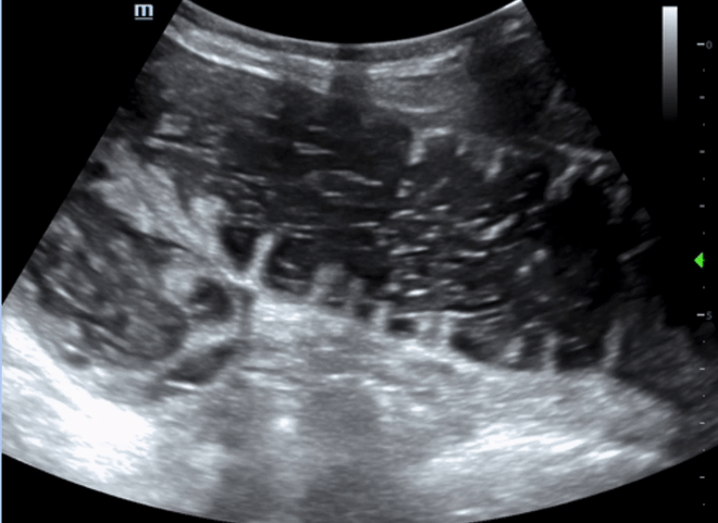
Bedside ultrasound of the abdomen shows:
Signs of SBO on US: Summary
Bowel diameter >/= 2.5 cm
Abnormal peristalsis:
To and fro movement
Keyboard sign (plicae circulares)
Confirms that you’re looking at small bowel (vs large bowel)
Bowel wall thickness >/= 3 mm
Extra-luminal fluid
1)Bowel size
The upper limit of normal diameter of the bowel is generally accepted as 3cm for the small bowel, 6cm for the colon and 9cm for the cecum (3/6/9 rule)

2)Abnormal Peristalsis
Abnormal peristalsis in SBO is generally described as “to-and-fro” with a shuttling or swirling appearance to the luminal contents.
Normal peristalsis

Abnormal to and fro peristalsis
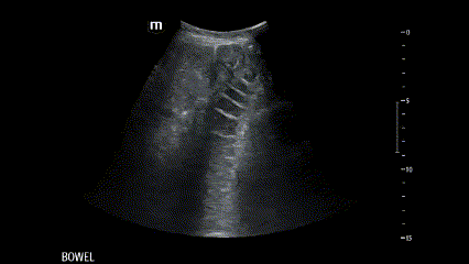
3)Keyboard sign
Visualization of the plicae circularis which is only present in small bowel, not large bowel
More clearly visible when the loops are spread out by liquid and when there’s bowel wall edema
Large bowel has haustra which are much further apart than plicae

In this same clip, notice the the finger like projections into the lumen (the plicae) that mark the small intestine
4)Bowel wall thickness
Normal adult bowel wall thickness is typically </= 3mm

Measurement 3 shows the thickened bowel wall
5)Extra-luminal fluid
Anechoic triangle between bowel loops = Collection of free fluid interspersed between bowel loops.
You can see in all the SBO clips above that there are triangles of fluid in between bowel loops.
Connotes high grade obstruction (and therefore worse prognosis)
High grade obstruction = complete obstruction, more likely to need surgical intervention

Extraluminal triangles of fluid
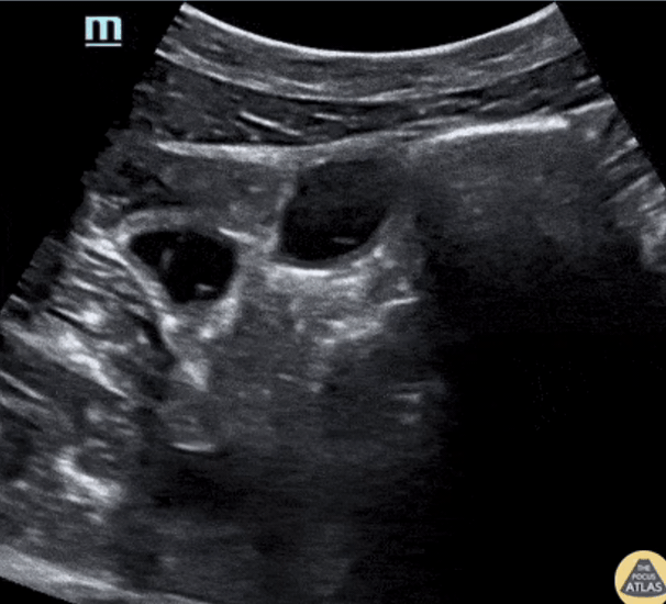
Here are some more clips from this patient:
Dilated small bowel loops see as soon as you place the probe on the patient

some more examples of extraluminal fluid, dilated bowel loops
Imaging for SBO
CT: Gold standard.
Sensitivities of 93%-96% and specificities of 93%-100% for the diagnosis of SBO
MRI: similar accuracy as CT, less accessible from the ED
Ultrasound: may be used as an adjunct to CT
Sensitivity 92.4% with 95% CI 89.0% to 94.7%) and specificity 96.6% with 95% CI 88.4% to 99.1% for SBO
Can be performed at bedside and interpreted immediately by the ED provider
No radiation
Accessible, fast
References:
Small bowel obstruction: Diagnosis by ultrasonography (aliem.com)