Distal Humerus Fractures
Written by, Suji Cha, DO; Edited by, Timothy Khowong, MH, MSEd
Anatomy:
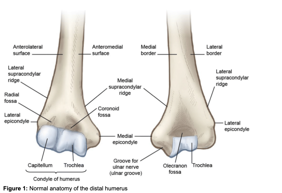
The distal humerus (figure 1) consists of the medial column, which consists of the medial epicondyle and trochlea and the lateral column, which consists of the lateral epicondyle and the capitellum. The capitellum and trochlea project anteriorly and together make up the condyle of the humerus. The capitellum articulates with the radial head and the trochlea articulates with the ulna to form the elbow joint.
Background:
Distal humerus fractures account for approximately 2% of all fractures in the adult population. These fractures include: supracondylar fractures, single column (condyle) fractures, bicolumnar fractures, and coronal shear fractures. There is a bimodal age distribution and is typically seen with high-energy trauma in young males or low-energy falls in elderly females. The mechanism of injury is typically a fall directly onto the elbow or onto an outstretched hand.
Classification:
There are three types of distal humerus fractures according to the AO/OTA Classification (figure 2):
Type A (Extra-articular)
Type B (Partial Articular)
Type C (Complete Articular)
Figure 2: Classification of distal humerus fractures
Assessment:
Patients will initially present with pain and swelling over the elbow with reduced range of motion secondary to pain. A thorough neurovascular exam is important, because the brachial artery, radial, ulnar, and median nerves are at risk of becoming injured from these fractures. It is also crucial to avoid assessing their range of motion due to risk of neurovascular injury. Monitor forearm compartments carefully as these patients may present with compartment syndrome.
Imaging:
Initial imaging includes plain radiographs with AP and lateral views of the elbow and humerus. Forearm imaging should be obtained as well to rule out coexisting injuries.
CT imaging is commonly obtained as it assists our orthopedics colleagues with surgical planning. It can also aid with assessing for possible shear fractures of the capitellum and/or trochlea, that are difficult to obtain without a true lateral view of the elbow on XR.
Management:
Treatment is almost always with an open reduction internal fixation surgery. Non-operative management with a cast for immobilization can be considered for high risk patients with significant comorbidities. Patients should be placed into a posterior long arm splint with a sling and referred to orthopedics surgery to discuss surgical treatment.
References:
1. Crean TE, Nallamothu SV. Distal Humerus Fractures. [Updated 2023 Jul 17]. In: StatPearls [Internet]. Treasure Island (FL): StatPearls Publishing; 2024 Jan-. Available from: https://www.ncbi.nlm.nih.gov/books/NBK531474/
2. Paras, Tyler, MD. Distal Humerus Fractures - Trauma - Orthobullets. www.orthobullets.com/trauma/1017/distal-humerus-fractures.
3. Lustosa L, Bell D, Double-arc sign. Reference article, Radiopaedia.org (Accessed on 04 Jun 2024) https://doi.org/10.53347/rID-148946
4. Distal Humerus Fractures of the Elbow - OrthoInfo - AAOS. orthoinfo.aaos.org/en/diseases--conditions/distal-humerus-fractures-of-the-elbow.


