
Ectopic POCUS
Written by: Dr. Rozalina Suleymanova
Edited by: Dr. Joann Hsu
The use of POCUS in female abdominal pain is vast:
Ovarian cysts
Ovarian torsion
Tubo-ovarian abscess
Uterine fibroids
IUD malplacement
Pathologic free fluid
Confirmation of IUP
Ectopic pregnancy
Let’s review some basic anatomy before we delve into ectopic pregnancy.
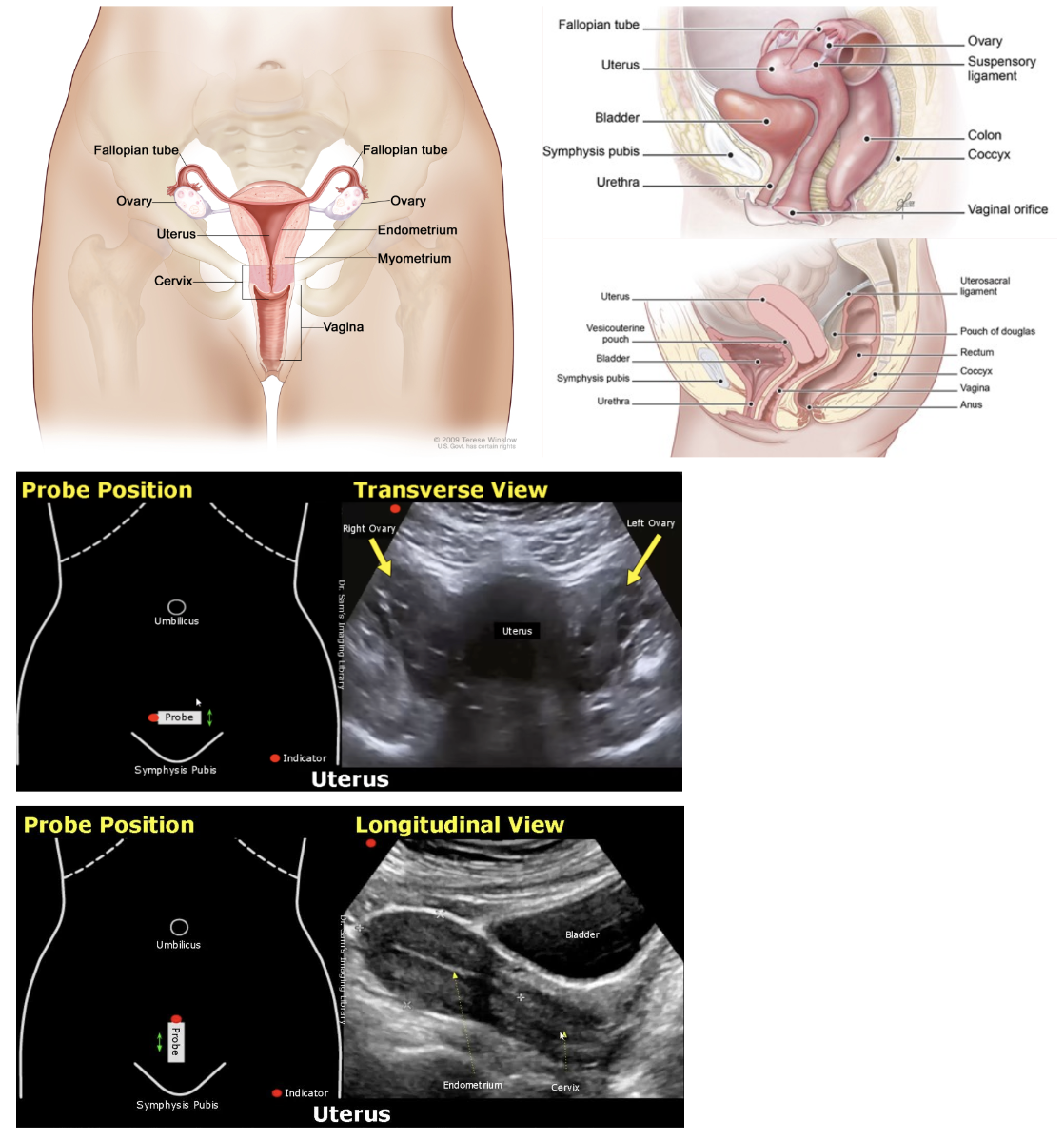
Case presentation:
34 year old female G4P2 with pmhx HTN who presented to ED with RLQ pain and vaginal bleeding x1 day. Abdominal pain began as intermittent cramping worse in the last day. Vaginal spotting started yesterday and was more severe this morning, prompting her visit to ED. Last sexually active 5 days ago. LMP 7/14/24 (one month ago). Takes progesterone only pill daily.
PERTINENT PE:
Patient is awake, alert, and in no acute distress
ABDOMEN AND GI: Soft; (+) suprapubic and RLQ tenderness, (-) guarding, (-) rebound, (-) Murphy's, (-) McBurney's, (-) CVA tenderness
Relevant labs: +HCG, HCG quantitative 3,028
Our bedside transabdominal pelvic ultrasound showed:
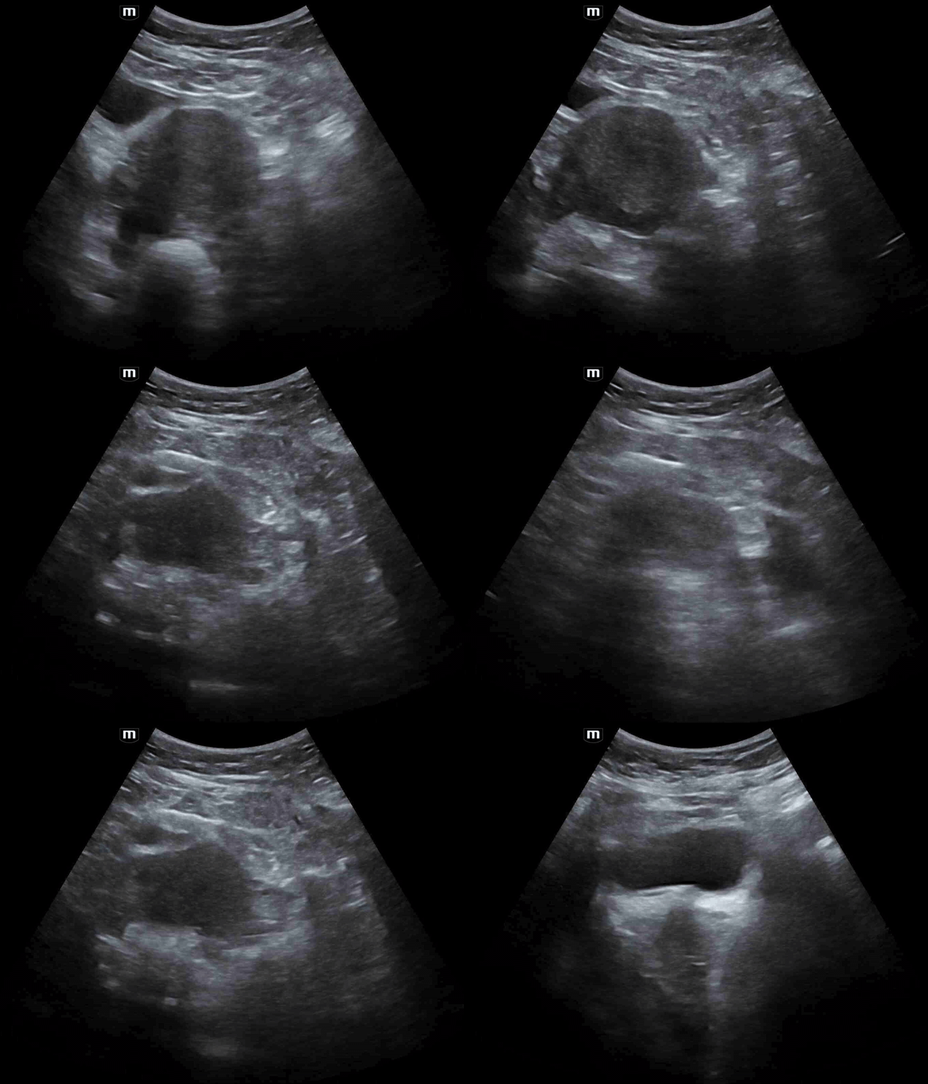
Transverse view of the uterus - there is NO IUP. Remember - the BHCG was positive.
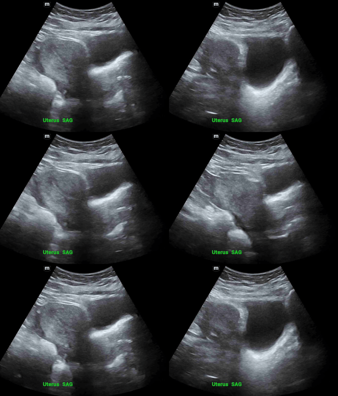
Sagittal view of the uterus again with no IUP and some free fluid
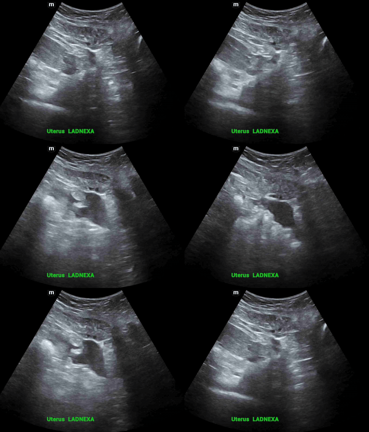
In the left adnexal area, there is a pocket of free fluid shown here.
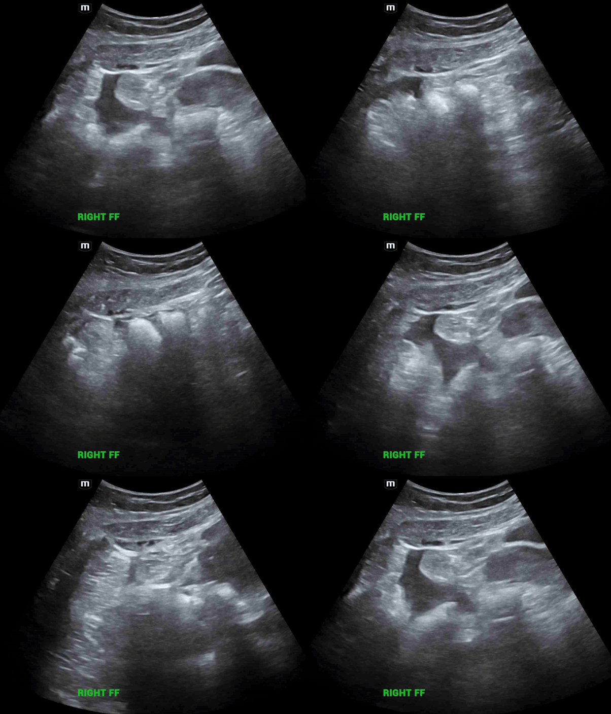
In the right adnexa, there is another pocket of free fluid.
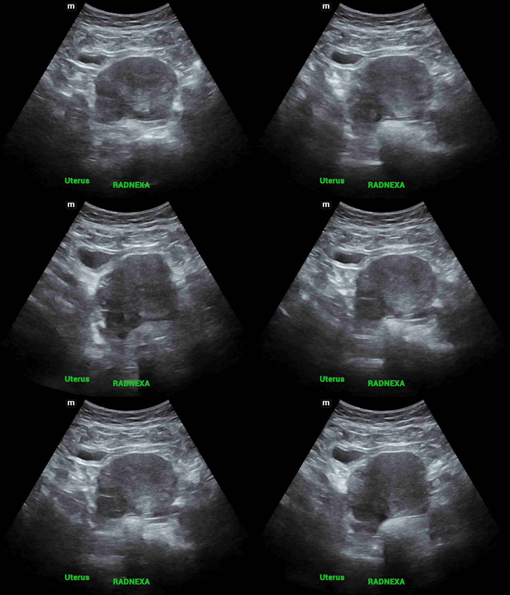
Looking globally at the right adnexal area, there is what appears to be a cyst-like structure that looks like an ovary next to the uterus.

Measuring this irregularly shaped structure containing a cyst. Unable to more clearly visualize this structure on transabdominal ultrasound.
So at this point, the patient should get a transvaginal ultrasound to better characterize the ovaries/adnexa.
Here are the patient’s official transvaginal ultrasound images:
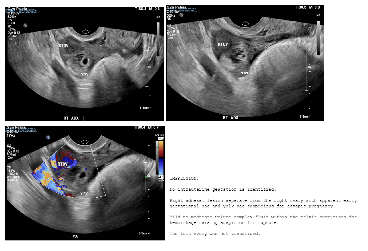
You can see more clearly that there is actually a structure separate from the right ovary that contains both a gestational sac and a yolk sac concerning for an ectopic pregnancy.
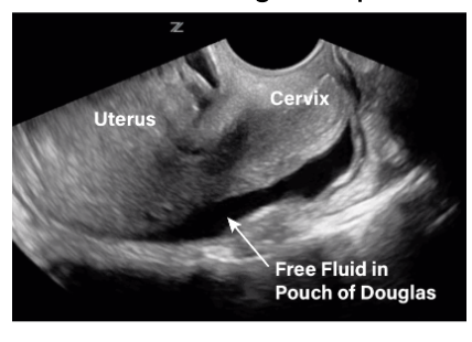
Free fluid in the pelvis
There can be a small amount of physiologic free fluid in women that is not necessarily pathologic, especially without symptoms such as pelvic pain.
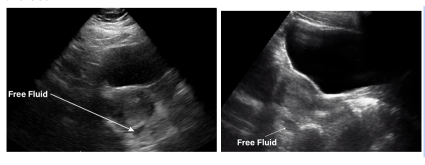
However there can be pathologic free fluid, which in the clinical context of ectopic pregnancy can be concerning for rupture of ectopic pregnancy.
A good estimation of amount of free fluid can be seen in the sagittal view. Above 1/3 the length of the sagittal uterus = significant free fluid, as shown below.
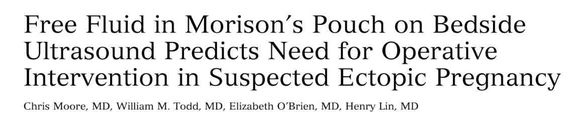
A recent prospective observational study investigated the relevance of finding free fluid in Morrison’s pouch (aka the RUQ view on the FAST exam) in women who had known ectopic pregnancies.
It was found that free fluid in Morrison’s pouch is highly specific (99.5%) for ectopic in the right clinical context and predicts the need for operative management.
TLDR: if concerned for ectopic, check the RUQ.
Doing so does not replace the transvaginal ultrasound study, but may be useful to support your diagnosis in low-resource community shops where there aren’t 24-hour ultrasound technicians.
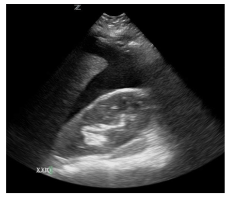
+ FF in the RUQ
Limitations of TAUS:
As with all POCUS, it’s operator dependent
Lower definition compared to TVUS (curvilinear vs endocavitary probe)
Views may be difficult to obtain in certain body habitus
Views may be obscured by abdominal content, eg. bowel gas
Patient disposition:
OBGYN was consulted and the patient was admitted for exploratory laparoscopy and removal of ectopic pregnancy. Ultimately she had a right salpingectomy and was discharged with 2 week post-op follow up.
Happy scanning!